Immune modulation plays a key role in regenerative medicine for multiple sclerosis (MS). At Stemedix, we focus on restoring immune balance to help reduce symptoms and slow disease progression. Regenerative medicine treatments, including stem cell therapies, target immune responses to decrease inflammation and support tissue repair. Since MS is an autoimmune condition, regulating immune function can help maintain quality of life and support overall health. According to the National Multiple Sclerosis Society, approximately 2.8 million people worldwide are living with MS, and around 1 million of those are in the United States. Effective immune modulation can help reduce relapses and manage symptoms, offering patients a better quality of life.
If you are considering regenerative medicine in Saint Petersburg, FL, Stemedix provides personalized treatment options designed to meet your needs. Our team is committed to guiding you through the potential benefits of regenerative medicine for MS, offering expert care every step of the way.
What is Immune Modulation?
Immune modulation is the process of adjusting the immune system’s response to either boost or suppress its activity, depending on the condition being treated. In regenerative medicine, it helps correct immune system imbalances in conditions like multiple sclerosis (MS). Instead of only addressing symptoms, this approach targets the underlying dysfunction. Regulating immune activity promotes balance, reduces inflammation, and supports tissue repair, offering a way to manage MS more effectively.
Immune System’s Role in Multiple Sclerosis
In multiple sclerosis (MS), the immune system wrongly attacks the myelin sheath that surrounds nerve fibers in the central nervous system. This causes nerve damage, inflammation, and a range of disabling symptoms. An estimated 85% of MS patients are initially diagnosed with relapsing-remitting MS (RRMS), which is characterized by clear relapses followed by periods of partial or complete recovery. Instead of protecting against harmful invaders, the immune system turns on the body’s own tissues.
Immune modulation through regenerative medicine works to correct this dysfunction by rebalancing the immune system, preventing further damage, and encouraging tissue repair. This approach not only alleviates symptoms but can also slow the progression of the disease, giving patients better chances for stability and improved function. By addressing the root cause, immune modulation helps the body heal naturally.
At Stemedix, we provide regenerative medicine in Saint Petersburg, FL, focusing on immune modulation to help manage MS. Our therapies aim to restore immune balance, promote tissue repair, and enhance your quality of life, offering a personalized path to long-term symptom relief and disease management.
The Science Behind Immune Modulation in Regenerative Medicine
Immune modulation in regenerative medicine often involves the use of stem cells, especially mesenchymal stem cells (MSCs). These cells help repair damaged tissues and regulate immune responses. In multiple sclerosis (MS), where the immune system attacks the body’s tissues, MSCs assist in restoring balance by reducing inflammation and encouraging tissue repair. This process helps prevent further immune attacks on the myelin sheath, providing relief and improving the overall condition of MS patients.
Stem Cells and Their Role in Immune Modulation
Mesenchymal stem cells (MSCs) have distinct characteristics that make them highly effective for immune modulation in multiple sclerosis (MS). They can release bioactive molecules that influence the immune system, reducing harmful immune responses and supporting tissue repair.
MSCs also reduce pro-inflammatory cytokines, which trigger inflammation, while promoting the activity of anti-inflammatory cells. This ability to balance the immune system and foster tissue regeneration makes stem cell therapy a vital component of regenerative medicine for MS.
For MS patients, stem cells not only help repair immune damage and restore balance but also ease symptoms like muscle pain, fatigue, and coordination problems. Instead of merely slowing disease progression, stem cell therapy provides a path to healing, improving overall health, and supporting long-term recovery.
Autologous vs. Allogeneic Stem Cell Therapy
In stem cell therapy for MS, there are two primary methods: autologous and allogeneic stem cell therapy. While each method offers unique benefits, both are designed to help modulate the immune system and promote healing.
Autologous Stem Cell Therapy: This approach uses the patient’s stem cells, which are collected and reintroduced into the body. Because these cells are from the patient, the risk of rejection is minimal, as the immune system typically recognizes them as “self.” However, the effectiveness may depend on the quality of the cells, especially in more advanced stages of the disease.
Allogeneic Stem Cell Therapy: Allogeneic stem cell therapy involves using stem cells from a donor. These cells are often more potent and can effectively modulate the immune system. They are also easily accessible, making them a good option for patients who cannot use their own cells. Although there is a slightly higher risk of immune rejection, improvements in stem cell processing have minimized this concern.
Both autologous and allogeneic stem cell therapies play an important role in regulating the immune system to treat MS. Each approach offers distinct benefits based on the patient’s specific condition, MS severity, and other health factors.
At Stemedix, we work closely with patients to determine the most suitable stem cell therapy based on their individual needs. Whether through autologous or allogeneic methods, we aim to use regenerative medicine treatments to restore immune balance, support healing, and enhance the quality of life for individuals living with multiple sclerosis.
How Immune Modulation Can Help Manage MS Symptoms
Immune modulation plays a key role in regenerative medicine treatments for multiple sclerosis (MS) by addressing the immune system dysfunction that causes the disease. Stem cell therapy and other immune-modulating treatments help restore immune balance, providing relief and slowing the progression of MS.
Slowing Disease Progression
Immune modulation plays a vital role in treating MS by slowing its progression. MS occurs when the immune system mistakenly attacks the myelin sheath, causing nerve damage and increased disability. Stem cell therapies, particularly mesenchymal stem cells, help regulate the immune response, reducing autoimmune attacks. This minimizes damage to the central nervous system and helps maintain nerve function.
By promoting tissue repair and supporting the body’s natural healing processes, stem cells reduce inflammation and prevent further deterioration. As a result, patients may experience fewer relapses and greater stability, leading to a better quality of life over time.
Reducing Inflammation
Inflammation is a key factor in the progression of MS symptoms, damaging the myelin sheath and causing issues like muscle spasms, pain, and cognitive difficulties. Stem cell therapy helps reduce inflammation by regulating the immune system, lowering pro-inflammatory cytokines, and activating anti-inflammatory cells.
By addressing the underlying cause of inflammation, stem cell therapy helps prevent further attacks on healthy tissue, reducing ongoing damage. Research indicates that MSCs can decrease levels of pro-inflammatory cytokines by up to 60%, significantly lowering inflammation and promoting tissue repair. This approach can ease symptoms such as muscle pain, spasticity, and neurological issues, ultimately improving mobility and lowering flare-up frequency. Many patients report notable relief, leading to an improved quality of life.
Symptom Control and Quality of Life
Immune modulation helps in controlling symptoms for MS patients by improving immune system function. Through regenerative medicine therapies, stem cells help address common MS symptoms such as muscle weakness, fatigue, and coordination issues. By restoring immune balance, these treatments prevent immune attacks that contribute to these symptoms, helping patients feel more energetic and in control.
As immune function improves, many patients notice an enhanced quality of life. With fewer symptoms, daily activities like walking, working, and spending time with loved ones become easier. This renewed independence can have a lasting positive impact, offering MS patients a better sense of well-being. Regenerative medicine supports individuals in regaining control over their health, enabling them to live more fully and manage their condition more effectively.
Why Choose Stemedix for Immune Modulation in MS Treatment?
Treating multiple sclerosis (MS) requires an approach that not only manages symptoms but also slows the progression of the disease. At Stemedix, we specialize in regenerative medicine in Saint Petersburg, FL, with a focus on immune modulation. Our therapies aim to address the underlying causes of MS while helping restore balance to the immune system.
Our Expertise in Regenerative Medicine
At Stemedix, we bring extensive experience and expertise in regenerative medicine, with a strong focus on stem cell therapies for autoimmune conditions like multiple sclerosis (MS). Our team is dedicated to using advanced stem cell science and immune modulation techniques to develop personalized treatment plans that address the unique needs of each patient. We recognize the challenges MS presents and its impact on the immune system, which is why our approach combines innovation with evidence-based practices.
We offer autologous stem cell therapies, utilizing the patient’s own cells to support healing and regeneration. Our experienced team conducts a thorough evaluation of each patient to create a personalized treatment plan tailored to their unique needs. By focusing on immune modulation, we aim to reduce inflammation, slow disease progression, and promote tissue repair, helping patients manage MS more effectively.
Patient-Centered Approach
At Stemedix, we prioritize our patients by offering a patient-centered approach to treatment. We understand that each individual’s experience with MS is different, which is why we tailor our care to fit your specific medical history, disease progression, and treatment goals.
From the moment you contact us, our dedicated care coordinators collaborate with you to create a personalized treatment plan. They are with you every step of the way, addressing questions, providing guidance, and offering support throughout your treatment. Whether it’s helping with travel arrangements, finding accommodations, or just offering reassurance, our care coordinators are committed to making your experience as seamless and comfortable as possible.
Positive Patient Outcomes
Choosing Stemedix for your immune modulation treatment can lead to positive results, as many patients with MS have reported improvements after stem cell therapy. A systematic review published by the National Institutes of Health reported that over 70% of MS patients treated with stem cell therapy experienced a reduction in relapses and improved mobility within six months of treatment. They have experienced relief from symptoms like muscle pain, inflammation, coordination challenges, and fatigue, which has helped enhance their overall well-being.
These positive results highlight the potential of immune modulation in managing MS. By targeting the root causes of immune system dysfunction, our treatments work to restore balance, reduce the severity of symptoms, and prevent additional neurological damage. This not only helps lower the frequency of MS flare-ups but also promotes better overall health and well-being.
The success stories from our patients demonstrate the effectiveness of our regenerative therapies, showing that Stemedix offers more than just treatment—we provide a path to a better quality of life. With a personalized approach, advanced therapies, and compassionate support, Stemedix is committed to helping you effectively manage MS.
Choosing Stemedix means choosing a treatment plan customized to your needs, supported by a team of experts who are dedicated to delivering the best possible care. We’re here to guide you through every step of your treatment journey, giving you the best opportunity to manage MS and improve your quality of life.
Stemedix: Harnessing Immune Modulation to Manage Multiple Sclerosis
Immune modulation plays an important role in managing multiple sclerosis (MS), giving patients the opportunity to improve how they cope with the disease. By targeting and regulating the immune system, this approach can help slow disease progression, decrease inflammation, and reduce symptoms that make everyday life challenging for those living with MS.
Stem cell therapies, a key aspect of regenerative medicine, offer a pathway to long-term relief by repairing damaged tissues and restoring balance to the immune system. This approach addresses the underlying cause of MS—autoimmune dysfunction—by modulating immune responses to reduce attacks on the central nervous system. As a result, MS patients often experience fewer flare-ups, reduced disability, and an overall enhancement in their quality of life.
By offering tangible improvements, immune modulation through regenerative medicine has become an essential treatment strategy in the fight against Multiple Sclerosis. Stemedix, based in Saint Petersburg, FL, leads the way in providing these specialty therapies, offering personalized treatment plans designed to meet each patient’s unique needs.
Take the first step toward managing MS effectively with Stemedix. Contact us at (727) 456-8968 or email us at yourjourney@stemedix.com to learn more about how our regenerative medicine treatments can help you.
Multiple sclerosis (MS) is a progressive neurological condition that affects millions of people worldwide. As this autoimmune disease disrupts the central nervous system, it leads to symptoms such as muscle weakness, numbness, and cognitive issues. In recent years, stem cell therapy has emerged as a promising treatment to alleviate these symptoms and potentially slow the progression of the disease.
At Stemedix, we recognize the challenges that MS patients face, particularly as the disease advances. We are dedicated to exploring stem cell treatments for multiple sclerosis as a potential solution. Stem cell therapy offers new hope by targeting the underlying causes of MS, especially the destruction of myelin—the protective sheath around nerve fibers. Myelin loss disrupts communication between the brain and the body, contributing to MS symptoms. Stem cells have the unique ability to regenerate damaged tissues, reduce inflammation, and modulate the immune system, which is critical in autoimmune diseases like MS.
Stem cell treatments for multiple sclerosis aim to restore function and slow the disease’s progression. Whether you’re experiencing early warning signs of multiple sclerosis, such as unexplained fatigue, numbness, or vision problems, or have been living with the disease for some time, stem cell therapy could offer a pathway to managing symptoms and improving your quality of life. In this article, we will explore how stem cell therapy for MS works, the scientific mechanisms behind it, and what you can expect from the treatment process. At Stemedix, we’re committed to helping you understand how stem cell treatments can make a difference in your journey with MS.
Stem Cell Therapy: A Game Changer for MS Treatment
Stem cell therapy offers a new approach to treating multiple sclerosis (MS), offering hope for many individuals living with this challenging condition. Understanding stem cells and their unique capabilities is essential in recognizing how stem cell therapy can be a powerful tool in MS treatment.
What Are Stem Cells?
Mesenchymal stem cells (MSC’s) are unique cells with the remarkable ability to transform into different cell types in the body. Known for their regenerative properties, they serve as the building blocks of life. In multiple sclerosis, stem cells can repair damaged tissues, including nerve cells affected by the disease. Unlike other cell types, stem cells are undifferentiated, meaning they can develop into specialized cells, such as those needed to regenerate the myelin sheath—the protective covering around nerve fibers often damaged in MS. While other treatments primarily manage symptoms or inflammation, stem cell therapy works to repair the underlying damage to the nervous system, making it a vital tool in regenerative medicine focused on healing rather than just symptom control.
Specialty Stem Cells in Multiple Sclerosis Treatment
Stem cell therapy for Multiple Sclerosis not only focuses on reducing inflammation but also on regenerating and repairing nerve damage. Certain specialized stem cells play an important role in this process:
Neural Stem Cells (NSCs): These cells have the potential to develop into various types of nerve cells, supporting the repair of damaged neurons and promoting neuroprotection. They may help restore function by replacing lost or injured nerve cells in MS patients.
Oligodendrocyte Precursor Cells (OPCs): Oligodendrocytes are responsible for producing myelin, the protective sheath around nerve fibers that is damaged in MS. Stem cell-derived OPCs aim to restore myelin, improving nerve function and slowing disease progression.
Schwann Cells: While primarily associated with the peripheral nervous system, Schwann cells play a role in myelin regeneration and nerve repair. Their regenerative properties make them an important consideration for supporting neural function in MS patients.
By incorporating these specialized stem cells into treatment strategies, regenerative medicine aims to go beyond symptom management and actively promote nerve repair and functional recovery. Stemedix continues to provide therapies informed by the latest research in stem cell applications for MS.
How Stem Cells Can Help MS Patients
Multiple sclerosis (MS) occurs when the immune system attacks the myelin, disrupting communication between the brain and the body. This leads to symptoms like numbness, muscle weakness, and cognitive challenges. Stem cells have the unique ability to regenerate the myelin sheath, repairing this damage. A key benefit of stem cells is their ability to reduce inflammation, which is central to the ongoing nerve damage in MS. By modulating the immune response, stem cells help control inflammation, providing symptom relief and potentially slowing disease progression. Stem cells may aid in regenerating damaged nerve cells and improving mobility, coordination, and cognitive function, making them a promising treatment option for MS.
At Stemedix, we recognize the challenges that come with MS, and we are committed to providing personalized stem cell treatments designed to address the root causes of the disease. Our goal is to offer a pathway to improved quality of life, aiming to slow the progression of MS and provide patients with the relief they need. If you’re considering stem cell therapy for MS, Stemedix is here to guide you every step of the way.
The Scientific Mechanisms Behind Stem Cell Treatments for MS
Stem cell therapy has become one of the most promising approaches to treating multiple sclerosis (MS). By targeting the underlying causes of the disease, stem cells offer a potential solution for repairing damage to the nervous system and improving overall function. Understanding the scientific mechanisms behind stem cell treatments can provide greater clarity on how these therapies work and why they hold so much potential for MS patients.
How Stem Cells Repair Damaged Myelin
Myelin is the protective covering around nerve fibers in the central nervous system, and its destruction is a key characteristic of multiple sclerosis (MS). When myelin is damaged, nerve signals cannot travel properly, resulting in symptoms like muscle weakness, numbness, and cognitive issues.
Stem cells can help regenerate myelin by transforming into oligodendrocyte precursor cells (OPCs), which produce new myelin. This regeneration improves nerve signal transmission and enhances overall function. Research, including animal models and early human trials, has shown promising results, with stem cell therapy leading to myelin repair and functional recovery. While still considered an emerging treatment, stem cell therapy’s potential to repair myelin offers hope for reducing MS symptoms and slowing disease progression.
Immune System Regulation
In multiple sclerosis, the immune system erroneously attacks myelin, causing progressive damage. Stem cells can modulate the immune system, reducing its overactive response and preventing further damage to the nervous system. This immune-modulating effect is critical in treating autoimmune conditions like MS.
Stem cells can reset the immune system by influencing T cells and B cells, which play a key role in attacking myelin. Ongoing research is investigating how stem cells can rebalance this immune response, potentially leading to long-term disease stabilization and fewer relapses. This immune modulation is a key mechanism of stem cell therapy for MS, addressing the disease’s root cause rather than merely managing its symptoms.
Reducing Inflammation and Enhancing Nerve Function
Chronic inflammation is another key feature of multiple sclerosis, contributing to the ongoing destruction of nerve cells and myelin. Stem cells can help combat this inflammation by producing anti-inflammatory cytokines, which are molecules that regulate the immune response. By reducing inflammation, stem cells help prevent further damage to the nervous system and support the body’s healing process.
Additionally, stem cells play a vital role in encouraging the repair of nerve cells and improving communication between the brain and the body. The regeneration of myelin and the reduction of inflammation work together to enhance nerve function, which can lead to improvements in mobility, coordination, cognitive function, and overall quality of life for MS patients.
Stem cell treatments for MS offer a multifaceted approach that addresses the damage caused by the disease, from repairing the myelin sheath to modulating the immune system and reducing inflammation. These scientific mechanisms provide a strong foundation for why stem cell therapy is considered a potential game-changer for those living with multiple sclerosis.
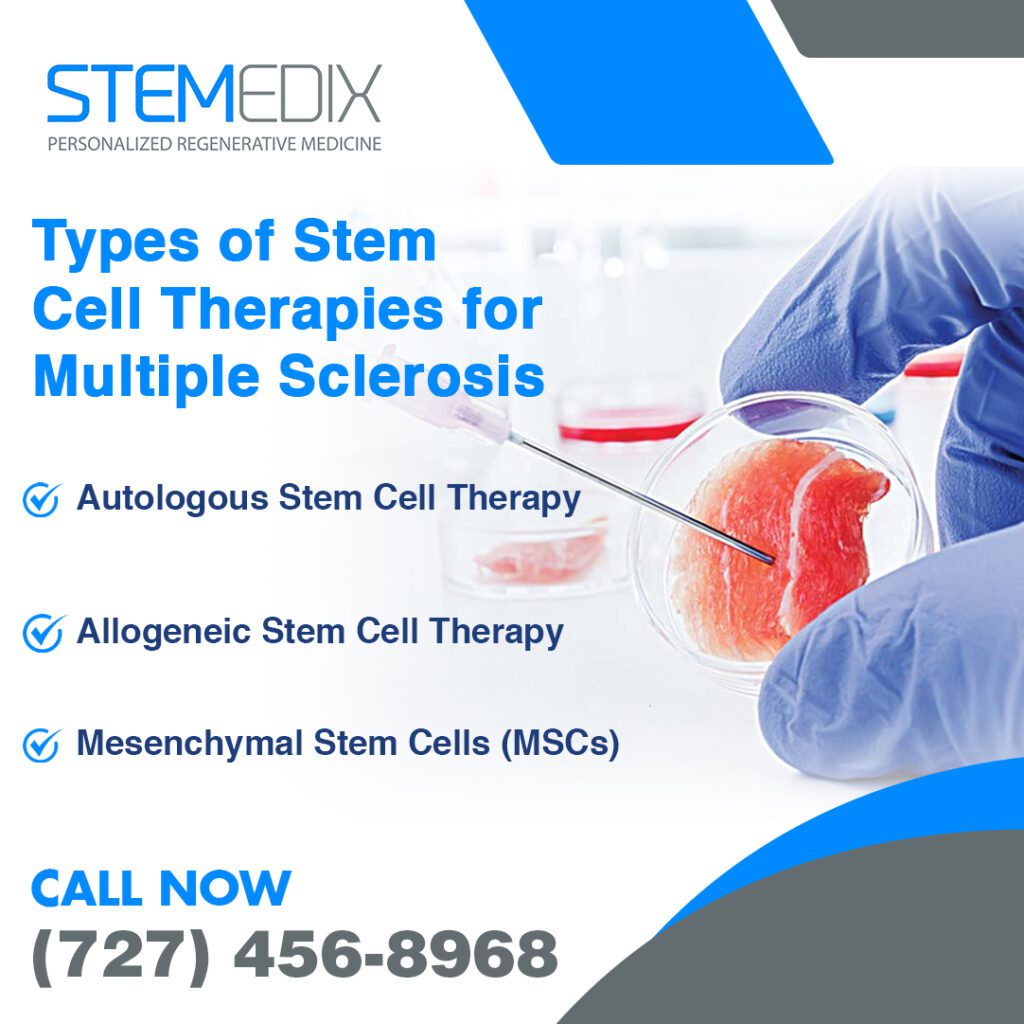
Types of Stem Cell Therapies for MS: Which One is Right for You?
Stem cell therapy is rapidly emerging as a viable option for individuals living with multiple sclerosis (MS). However, there are different types of stem cell therapies, each with unique processes and potential benefits. Understanding the different options available can help you make an informed decision about the treatment that’s best for you.
Autologous Stem Cell Therapy
Autologous stem cell therapy uses the patient’s own stem cells, offering a highly personalized treatment for multiple sclerosis (MS). The process begins with collecting stem cells from the patient’s bone marrow or blood. These cells are then purified in a laboratory and reintroduced into the body to help regenerate damaged tissues, repair myelin, and modulate the immune system.
A significant benefit of autologous stem cell therapy is the elimination of immune rejection, as the cells are derived from the patient’s own body. This reduces complications associated with foreign tissue. However, challenges include the time-consuming, expensive nature of the process and limited stem cell availability in some patients, especially older individuals. Despite these hurdles, it remains a popular and effective MS treatment.
Allogeneic Stem Cell Therapy
Allogeneic stem cell therapy uses stem cells from a healthy donor rather than the patient’s own cells. These donor cells are harvested, processed in a lab, and transplanted into the patient. This approach is helpful when a patient’s stem cells are not viable or when a quicker stem cell replenishment is needed.
One key benefit is the immediate availability of high-quality donor cells that can regenerate tissue, repair myelin, and modulate the immune response in MS patients.
Mesenchymal Stem Cells (MSCs)
Mesenchymal stem cells (MSCs), typically sourced from umbilical cord tissue (UCT), adipose tissue, or bone marrow, hold significant promise for treating multiple sclerosis (MS). These cells are known for reducing inflammation, promoting tissue repair, and aiding in the regeneration of damaged myelin. MSCs also modulate the immune system, addressing the autoimmune response driving MS progression.
MSC therapy has garnered attention for its potential to repair MS-related damage while addressing immune dysfunction. These cells release anti-inflammatory cytokines, alleviating chronic inflammation. Additionally, MSCs may aid in nerve tissue repair, improving mobility and cognitive function. While research is ongoing, early findings suggest MSC therapy could reduce relapses, manage symptoms, and even slow disease progression, enhancing the quality of life for MS patients.
At Stemedix, we offer a range of stem cell treatment options tailored to your individual needs. Our team of experts can help you determine the most suitable approach for managing your MS. We’re committed to providing advanced treatments that allow you to live a better life with MS, and our personalized care guarantees that you receive the best possible outcomes.
What Does the Stem Cell Treatment Process Involve for MS?
Stem cell therapy is an evolving treatment option for multiple sclerosis (MS), offering hope for patients seeking ways to manage their symptoms and slow disease progression. Understanding the stem cell treatment process is essential for anyone considering this approach. Here’s a detailed look at what you can expect throughout the process, from your initial consultation to the post-treatment phase.
Initial Consultation and Patient Evaluation
The initial step in the stem cell treatment process for MS is the consultation with a healthcare provider. During this meeting, the provider will review your medical history, conduct a thorough examination, and evaluate any early warning signs of multiple sclerosis, such as unexplained fatigue, numbness, or vision problems.
Diagnostic tests, including MRI scans and blood tests, may be recommended to evaluate the extent of myelin damage and inflammation. Based on these results, the provider will discuss different stem cell therapy options. This guarantees a personalized treatment plan that aligns with your medical history and the progression of MS, guiding you toward the most suitable approach.
Stem Cell Collection and Processing
Once the type of stem cell therapy is determined, the next step is stem cell collection. For autologous therapy (using your own cells), stem cells are typically harvested from your bone marrow or adipose (fat tissue). In the case of allogeneic therapy (using donor cells), stem cells are sourced from a carefully screened donor to make sure compatibility.
After collection, the stem cells are processed in a laboratory where they are isolated, purified, and prepared for reintroduction into the body. This step is essential to make sure that the cells are viable and effective. For mesenchymal stem cells (MSCs), special techniques are employed to enhance their ability to repair tissue, reduce inflammation, and regenerate damaged myelin.
Injection and Treatment Procedures
Once the stem cells are prepared, they are reintroduced into your body. Depending on the therapy type, this may be done through an intravenous infusion or direct injections into affected areas, such as the spinal cord or regions with significant nerve damage. This approach targets areas that need repair.
The treatment duration varies based on the selected therapy and individual patient needs. Some treatments may take a few hours, while others require multiple sessions over weeks or months. Throughout the process, your healthcare provider will closely monitor progress, including improvements in mobility, muscle strength, and cognitive function, and adjust the treatment plan as needed to achieve the best possible outcome.
Tracking Progress and Long-Term Care
After the treatment, regular follow-up appointments are vital for tracking your progress. Your healthcare provider will continue to monitor your response to stem cell therapy, which may include conducting tests to evaluate changes in symptoms and overall function. This allows for adjustments to the treatment plan as necessary to guarantee continued progress in managing MS.
At Stemedix, we understand that each patient’s journey with multiple sclerosis is unique. Our experienced team is committed to providing personalized care throughout every stage of the stem cell therapy process. We work closely with you to get the best possible outcome and offer ongoing support as you traverse the challenges of living with MS.
Stem cell therapy offers a promising path forward for many people with multiple sclerosis. By partnering with healthcare providers who specialize in these advanced treatments, you can explore the potential benefits and make informed decisions about your health and well-being.
Why Choose Stemedix for Stem Cell Therapy for MS?
When considering stem cell treatments for multiple sclerosis (MS), selecting the right provider is very important to ensuring the best possible outcomes. At Stemedix, we specialize in offering advanced regenerative treatments that are personalized to each patient’s specific needs. Our commitment to delivering exceptional care and effective stem cell therapies for MS is backed by years of expertise in treating neurodegenerative diseases, including multiple sclerosis.
Expertise in Stem Cell Treatments
At Stemedix, we have a proven track record of success in treating multiple sclerosis and other neurodegenerative conditions with stem cell therapy for MS. Our team brings extensive experience and knowledge to each treatment plan, ensuring that you receive the most effective care for your unique situation.
What sets us apart is our ability to combine scientific advancements with personalized care. We understand that MS affects each individual differently, which is why we tailor our treatment plans to address your specific symptoms, disease progression, and overall health. Our specialists are well-versed in the latest stem cell therapies, including autologous and allogeneic stem cell options. They will work closely with you to choose the most appropriate therapy for your needs.
Supportive Care Throughout the Treatment Process
Going through the complexities of MS and stem cell therapy can be overwhelming, but with Stemedix, you’ll never feel alone. From the moment you reach out for a consultation, our team of care coordinators will be there to support you every step of the way. Whether you need assistance with scheduling, understanding the treatment process, or managing the emotional aspects of your journey, we are here to make sure that you feel informed, comfortable, and confident throughout your experience.
We offer continuous support before, during, and after your stem cell treatment. This is especially important for MS patients, who may need additional assistance to track progress and manage any challenges during recovery. Our care coordinators are dedicated to guiding you through the process, offering consistent follow-up, and making sure that you feel empowered in your healthcare decisions.
Stemedix: A New Hope for Patients with MS
Stem cell therapy has emerged as a promising treatment option for multiple sclerosis (MS), offering hope to those living with this challenging condition. As we’ve discussed, stem cells have the potential to repair the damage caused by MS, particularly by regenerating myelin, reducing inflammation, and modulating the immune system. Unlike traditional treatments, stem cell therapy addresses the underlying causes of MS, which can lead to more effective management of symptoms. By stimulating the body’s natural regenerative processes, stem cells may help improve nerve function and slow the disease’s progression. If you’ve noticed early warning signs of multiple sclerosis, such as unexplained fatigue, numbness, or vision problems, stem cell therapy could offer a potential solution.
For MS patients, stem cell therapy can offer significant benefits, including better mobility, improved cognitive function, and enhanced overall quality of life. Though research continues to evolve, the results so far suggest that stem cell therapy could be a valuable tool for managing MS symptoms more effectively. If you’re living with MS and want to explore new treatment options, stem cell therapy could be the solution you’ve been searching for. At Stemedix, based in Saint Petersburg, FL, we offer personalized care and advanced stem cell treatments designed to help you manage your MS symptoms and improve your quality of life. Our team is here to support you from the initial consultation to post-treatment care.
Contact Stemedix today at (727) 456-8968 or email us at yourjourney@stemedix.com to schedule your consultation. Let us help you discover how stem cell therapy can make a difference in your journey with MS.
Parkinson’s disease is a progressive neurological disorder that can significantly affect daily life. Understanding its early signs and seeking a timely diagnosis can make a crucial difference in managing this condition. At Stemedix, we recognize the importance of being informed about the symptoms, diagnostic approaches, and available treatments, including the promising field of stem cell therapy. Our goal is to empower you with knowledge, guide you through the complexities of Parkinson’s disease, and discuss the potential of emerging treatments like stem cell regenerative therapy as a complementary option in managing symptoms. By staying vigilant about the early signs, you can take proactive steps toward better health and well-being.
Overview of Parkinson’s Disease
Parkinson’s disease is a neurological condition that mostly affects movement and is complicated and progressing. Understanding Parkinson’s begins with recognizing that it is classified as a movement disorder. This condition stems from the degeneration of specific nerve cells in the brain, significantly impacting your body’s ability to control movements effectively.
Symptoms typically develop gradually and may begin with subtle changes in your daily activities. You might notice a slight tremor in your hand or a change in your walking pattern. As the disease progresses, these symptoms can become more pronounced, leading to difficulties with balance, coordination, and overall motor function. Beyond physical movement, Parkinson’s can also affect emotional and cognitive aspects of life, highlighting its widespread impact on daily living.
The emotional weight of receiving a Parkinson’s diagnosis can be heavy. It’s important to know you are not alone in this journey. Millions of people are going through similar challenges, and there are communities and resources available to support you. Moreover, the exploration of various treatment options, including innovative therapies like stem cell regenerative therapy, is continuously evolving. This progress gives hope to those affected by the disease.
Pathophysiology of Parkinson’s Disease
In Parkinson’s disease, there is a significant change in the brain’s structure and function. Dopamine-producing neurons in the substantia nigra, a part of the brain, gradually die off as part of the pathophysiology. Dopamine is an essential neurotransmitter that aids in controlling emotions and actions. When the neurons that produce dopamine begin to deteriorate, the balance of neurotransmitters in your brain becomes disrupted, leading to the hallmark symptoms of Parkinson’s.
As dopamine levels decrease, you may experience a range of motor symptoms. These can include tremors, stiffness, slowness of movement, and impaired balance. Each person’s experience can vary widely, making it essential for you to pay attention to your unique symptoms and communicate them with your healthcare provider. Understanding the underlying changes in your brain can empower you to engage actively in your treatment and management options.
The loss of dopamine-producing neurons also sheds light on some non-motor symptoms that are often overlooked. These may include changes in mood, sleep disturbances, and even cognitive decline. Recognizing these aspects is crucial for creating an overall management plan.
At Stemedix, we focus on an individualized approach that considers not just the motor symptoms but also the overall well-being of our patients. By understanding the full scope of Parkinson’s disease, you can take a proactive stance in your journey toward improved health and quality of life.
Early Symptoms of Parkinson’s Disease
Motor Symptoms: Recognizing the First Signs
When it comes to Parkinson’s disease, early recognition of motor symptoms can be pivotal. The initial signs are often subtle and may be dismissed as normal signs of aging or fatigue. One of the most common early symptoms you might notice is a tremor, typically starting in the hand or fingers. This involuntary shaking can occur when the hand is at rest and may be more pronounced during periods of anxiety or stress.
Stiffness is another hallmark symptom that can creep in gradually. You may find that your muscles feel rigid, making it difficult to do everyday activities like buttoning a shirt or reaching for objects. This rigidity can also affect your posture and lead to a stooped stance.
Bradykinesia, or slowness of movement, often becomes noticeable as well. You might experience a decrease in your overall speed when walking or performing movements, which can become frustrating and impact your daily activities. Recognizing these motor symptoms early on can be vital for initiating treatment and management strategies, allowing for a better quality of life.
Non-Motor Symptoms: The Hidden Indicators
While motor symptoms tend to grab attention, it is crucial not to overlook the non-motor symptoms that can signal the onset of Parkinson’s disease. You may experience cognitive changes, such as difficulty concentrating or a decline in memory. These cognitive shifts can be concerning and may affect your ability to manage day-to-day responsibilities.
Emotional changes are also significant indicators. Feelings of anxiety or depression can emerge early in the disease and may not be immediately associated with Parkinson’s. It’s essential to understand that these emotional responses are a natural reaction to the changes occurring within your brain and body.
Sensory changes, such as altered sense of smell or changes in vision, can also occur. You might notice a reduced ability to detect odors or a decrease in visual acuity. By identifying these non-motor symptoms, you and your healthcare practitioner may develop a thorough treatment plan that takes into account the disease’s emotional and physical components.
The Importance of Early Recognition
Recognizing the early symptoms of Parkinson’s disease, both motor and non-motor, is critical for several reasons. Firstly, early identification allows for timely intervention, which can lead to improved management of the disease. If you notice symptoms like tremors, stiffness, or changes in mood, it’s essential to consult with your healthcare provider. Early diagnosis can facilitate more effective treatment strategies, including medication and lifestyle adjustments.
Furthermore, being proactive about recognizing symptoms can empower you to take control of your health journey. Engaging in early treatment can help mitigate the progression of symptoms and enhance your overall quality of life. At Stemedix, we believe in a patient-centered approach that emphasizes the importance of awareness and early intervention. By understanding your body and its signals, you can go through this journey more effectively, potentially exploring advanced treatment options such as stem cell regenerative therapy as part of your management strategy.
Diagnostic Approach to Parkinson’s Disease
Initial Patient Evaluation
The journey to a Parkinson’s disease diagnosis starts with an initial evaluation by healthcare professionals. This step is crucial for understanding your symptoms and medical history. During this evaluation, your provider will ask detailed questions about your symptoms, their onset, and how they’ve progressed. You may discuss specific motor symptoms, like tremors or stiffness, alongside any non-motor symptoms, such as mood changes or cognitive issues.
A comprehensive medical history is equally important, as it may reveal genetic predispositions or environmental factors. In some cases, a referral to a neurologist specializing in nervous system disorders will occur. Your input during this evaluation is invaluable; being open and detailed will enable healthcare professionals to make informed assessments and create an effective management plan.
Diagnostic Criteria for Parkinson’s Disease
Once the initial evaluation is complete, healthcare professionals will use specific diagnostic criteria to confirm a diagnosis of Parkinson’s disease. The most widely used set of criteria comes from the movement disorder society-unified Parkinson’s Disease rating scale (MDS-UPDRS). This scale includes several components that assess various aspects of the disease.
The MDS-UPDRS evaluates motor functions, non-motor experiences, and daily living activities affected by Parkinson’s. Healthcare providers will look for key signs, such as bradykinesia, rigidity, and postural instability. A combination of these symptoms, particularly when they are present alongside characteristic tremors, can help solidify the diagnosis.
It’s important to understand that no single test can confirm Parkinson’s disease. Instead, the diagnosis is often based on clinical observation and the presence of specific symptoms over time. Engaging in an open dialogue with your healthcare provider about your symptoms will support accurate diagnosis and help you understand the rationale behind their assessments.
Diagnostic Imaging and Tests
In addition to clinical evaluations, various imaging techniques and tests can aid in diagnosing Parkinson’s disease. While these tools cannot definitively confirm the condition, they help rule out other neurological disorders that may present with similar symptoms. Magnetic resonance imaging (MRI) is often used to examine the brain’s structure, identifying signs of other conditions, such as strokes or tumors, that might mimic Parkinson’s symptoms. Positron emission tomography (PET) scans provide insights into brain function by measuring neuronal metabolic activity. These scans visualize dopamine production and reveal abnormalities linked to Parkinson’s disease.
Additional tests, like blood tests or assessments of olfactory function, can provide further support. Ultimately, your healthcare team will combine clinical evaluations, diagnostic criteria, and imaging results to form a comprehensive diagnosis.
At Stemedix, we understand that navigating the diagnostic process can be overwhelming. You can actively participate in your health journey by encouraging open communication with your healthcare professional and being proactive in talking about your symptoms. Developing a management strategy that may incorporate cutting-edge therapeutic alternatives like stem cell regeneration therapy requires an early and precise diagnosis.
Investigating Stem Cell Therapy for Parkinson’s Disease
Mechanism of Action: How Stem Cells Can Help
Stem cell therapy represents a novel approach to treating Parkinson’s disease, aiming to address the underlying neurological damage that characterizes the condition. The potential of stem cells is seen in their capacity to repair damaged neurons and give the afflicted brain regions their normal function. The hallmark symptoms of Parkinson’s disease, such as tremors and rigidity, are linked to the loss of dopamine-producing neurons. Stem cells are being explored for their potential to support brain repair and reduce symptom severity, though definitive reversal remains a goal for future research.
When administered, stem cells may have the potential to differentiate into neuron-like cells and support neuronal health. Researchers are investigating whether these cells can indirectly aid in restoring dopamine production and improving motor functions. The goal of stem cell therapy for Parkinson’s is not only to alleviate symptoms but also to modify the disease’s progression by repairing the neurological pathways involved.
At Stemedix, we focus on exploring advanced techniques in stem cell regenerative therapy, which are under investigation as potential tools for managing symptoms and improving the quality of life in individuals with Parkinson’s disease. Our approach emphasizes not just treatment but an extensive understanding of how stem cells can work within the body to promote healing and recovery.
Promising Research Findings
Recent studies have indicated the potential benefits of stem cell therapy for Parkinson’s patients in experimental settings. While some participants reported improvements in motor function and quality of life, these results are still under investigation, and more rigorous clinical trials are needed to establish effectiveness and safety. These studies highlight the potential for stem cells to help restore neuronal health, addressing the underlying damage caused by the disease and enhancing the overall functioning of the nervous system.
Preliminary studies have reported some participants experiencing improvements in motor symptoms, such as tremors and dexterity, following stem cell therapy. However, outcomes vary significantly, and further research is needed to confirm these findings and understand long-term effects.
Moreover, ongoing research is exploring various types of stem cells, including mesenchymal stem cells, which have shown promise in modulating inflammation and supporting neuroprotection in the brain. This exciting field of study continues to evolve, with clinical trials underway to further investigate the long-term efficacy and safety of stem cell therapy for Parkinson’s disease.
At Stemedix, we stay abreast of these developments, integrating the latest findings into our patient care practices. By leveraging advanced research, we aim to offer our patients the best possible outcomes through innovative stem cell regenerative therapy.
Stemedix’s Role in Stem Cell Therapy for Parkinson’s Disease
At Stemedix, we are committed to providing personalized treatment options for individuals with Parkinson’s disease, utilizing stem cell regenerative therapy to potentially enhance patient care and improve quality of life. Our approach begins with a comprehensive evaluation of each patient’s unique condition, allowing us to tailor treatments that address their specific needs and health goals.
We understand that navigating the complexities of Parkinson’s disease can be overwhelming, which is why our dedicated team of healthcare professionals is here to guide you every step of the way. From initial consultations to ongoing support, we emphasize compassionate care and patient education, ensuring you have the information and resources needed to make informed decisions about your health.
Through our stem cell therapy programs, we harness the potential of regenerative medicine to help promote healing and recovery. By employing progressive techniques, we strive to optimize the therapeutic benefits of stem cell therapy, aiming to restore function and enhance the overall quality of life for our patients.
At Stemedix, we believe in a future where individuals with Parkinson’s disease can achieve improved health outcomes and live fulfilling lives. Our commitment to innovation and excellence in patient care sets us apart as a leader in the field of regenerative medicine, and we are excited to be part of your journey toward wellness.
Future Prospects of Stem Cell Therapy in Treating Parkinson’s Disease
Advances in Stem Cell Research
The field of stem cell research is rapidly evolving, especially concerning its applications in treating Parkinson’s disease. Current clinical trials are underway, focusing on various aspects of stem cell therapy, including the types of stem cells used, delivery methods, and patient outcomes. These studies aim to determine the most effective ways to utilize stem cells to restore neuronal function and alleviate the symptoms associated with Parkinson’s.
Researchers are exploring various sources of stem cells, such as induced pluripotent stem cells (iPSCs) and mesenchymal stem cells, each offering unique benefits and challenges. For instance, iPSCs are particularly exciting because they can be generated from a patient’s own cells, potentially reducing the risk of immune rejection. Ongoing trials are examining not only the efficacy of these therapies but also the timing of treatment, as an earlier intervention may yield better results in terms of neuroprotection and functional recovery.
At Stemedix, we are closely monitoring these advancements and integrating promising findings into our treatment protocols. By participating in research collaborations and keeping our finger on the pulse of new developments, we aim to provide our patients with pioneering options that could significantly improve their health outcomes.
Safety Considerations and Ethical Implications
As we explore the potential of stem cell therapy for Parkinson’s disease, it is crucial to address the safety considerations and ethical implications of experimental treatments. Many therapies remain unproven and are only available through regulated clinical trials under the oversight of organizations like the FDA, which ensures rigorous safety and efficacy standards.
Ethical considerations also play a vital role in the advancement of stem cell therapy. The sources of stem cells raise important ethical questions regarding consent, sourcing, and potential commercialization.
At Stemedix, we adhere to strict ethical guidelines and practices, ensuring that all our procedures are conducted with transparency and respect for patient autonomy. We prioritize informed consent and actively engage our patients in discussions about the ethical dimensions of their treatment options.
Accessibility of Emerging Treatments
The future landscape of treatment options for patients with Parkinson’s disease is promising, with a growing emphasis on making innovative therapies more accessible. As research progresses and more evidence supports the effectiveness of stem cell therapy for Parkinson patients, we anticipate an increase in treatment centers offering these options. This expansion can help reduce disparities in access to care, ensuring that more individuals benefit from the potential improvements that stem cell therapy can provide.
At Stemedix, we are dedicated to enhancing accessibility for our patients by providing personalized treatment plans that fit individual needs and circumstances. We understand that navigating the healthcare system can be challenging, especially when seeking advanced therapies. Our team is here to assist you through every step, from initial consultations to ongoing support throughout the treatment journey.
As we move forward, the integration of stem cell therapy into the treatment landscape for Parkinson’s disease holds the potential to transform patient care. By focusing on research, ethical practices, and accessibility, we at Stemedix aim to be at the forefront of these developments and are committed to improving the lives of Parkinson’s patients.
Empowering Lives with Stemedix – Navigating the Journey of Parkinson’s Disease
The journey through Parkinson’s disease can be daunting, but understanding the significance of early diagnosis plays a crucial role in improving outcomes for persons affected by this condition. Recognizing the initial symptoms, whether they are motor or non-motor, allows for timely intervention and management, which can significantly enhance the quality of life.
As we look to the future, the promise of stem cell therapy offers new hope. This innovative approach not only aims to address the underlying neurological damage caused by the disease but also holds the potential to restore function and improve patient well-being.
At Stemedix, located in Saint Petersburg, FL, we are dedicated to harnessing the power of stem cell regenerative therapy to provide personalized treatment options that meet the unique needs of our patients. By prioritizing research and ethical practices, we strive to be a leader in advancing care for those with Parkinson’s disease.
If you or a loved one is experiencing symptoms of Parkinson’s disease, early diagnosis and tailored treatment options are crucial. At Stemedix, we specialize in personalized care and stem cell therapy. Contact us today at (727) 456-8968 to learn more about your treatment options.
If you’ve been experiencing hair loss, you may be considering your treatment options. Many individuals have little success with widely available products that promise hair regrowth, and understandably want to avoid invasive procedures such as hair transplantation. Fortunately, there’s another option: stem cell hair restoration.
What Is Stem Cell Hair Restoration?
Stem cell therapy for hair restoration employs the use of stem cell exosomes, powerful components with an important role in cellular communication. They have over 200 signaling proteins and can turn on regenerative factors, prompting the cellular survival that can help to prevent further hair loss and stimulate new growth. Patients often notice new hair growth after just one session, with additional treatments leading to even better results.
How Much Does it Cost?
Stemedix offers stem cell therapy for hair restoration starting at $3,900. When you consider the cost of hair transplant surgeries, which can vary from $4,000 to $15,000, stem cell hair treatments become an attractive, cost-effective option. This is especially true considering the fact that, as with any surgery, recovery from hair transplants may come with additional costs, including pain medications and the cost of treating infections (the most common complication of the treatment).
Moreover, you simply can’t put a price tag on the boosted confidence, comfort, and overall improved quality of life that comes with thicker, fuller hair. Whether you’ve dodged social events or you’re experiencing low self-esteem, stem cell hair restoration could be the solution that allows you to feel like yourself once again. Hair loss has even been linked to higher levels of stress, anxiety, depression, and social phobias. Thus, finding an effective solution to restore your hair growth is about much more than cosmetics alone, and it could be the very factor that allows you to embrace a new chapter of your life.
If you find yourself noticing early signs of hair loss and you would like to reap the benefits of preventive stem cell measures contact us today for more information!
For millions of people across the globe, orthopedic issues cause discomfort, limited mobility, and other frustrating symptoms which make it challenging to participate in daily activities. Osteoarthritis, for example, affects the joints by wearing away the cushiony cartilage between bones. While the prevalence of the condition is on the rise, conventional treatments are still lacking: patients are often required to choose between temporary fixes such as medications to mask the pain or invasive surgical procedures. And, this isn’t isolated to osteoarthritis; patients with other orthopedic issues face similar dilemmas. For those suffering, there are several benefits of stem cell therapy for orthopedic issues.
This treatment involves the targeted administration of stem cells, which can self-renew and transform into virtually any cell type in the body. Researchers believe they hold tremendous healing potential for orthopedic issues due to their abilities to change into cartilage cells, minimize inflammation, and release cytokines to reduce pain and slow the degenerative process.
Below, discover some of the benefits of stem cell therapy for orthopedic issues, as well as the types of conditions they can treat.
Common Orthopedic Conditions Treated with Stem Cell Therapy
The potential applications for stem cell therapy span far and wide. Not only have they been used to treat orthopedic conditions, but they’re also used for patients with neurodegenerative and autoimmune conditions.
Some of the specific orthopedic issues being treated by stem cell therapy include:
· Osteoarthritis
· Sports and athletic injuries
· Musculoskeletal injuries
· Spinal cord injuries
· Degenerative disc disease
Benefits of Stem Cell Therapy for Orthopedic Issues
There are many advantages to stem cell therapy. Here are just a few of the most noteworthy benefits to consider:
· Reduced pain and inflammation
· The ability to delay or prevent the need for invasive surgery
· Increased healing potential
· An all-natural treatment
· Safe and effective
Finally, but perhaps most importantly, this regenerative therapy can give new hope to patients who haven’t seen results from other treatment methods. Stem cell treatment could help to restore mobility, alleviate pain, reduce inflammation, and enhance overall quality of life — outcomes which may have previously seemed unattainable to patients with orthopedic issues.
This website and its contents are not intended to treat, cure, diagnose, or prevent any disease. Stemedix, Inc. shall not be held liable for the medical claims made by patient testimonials or videos. They are not to be viewed as a guarantee for each individual. The efficacy for some products presented have not been confirmed by the Food and Drug Administration (FDA).
This website uses cookies to improve your experience while you navigate through the website. Out of these cookies, the cookies that are categorized as necessary are stored on your browser as they are essential for the working of basic functionalities of the website. We also use third-party cookies that help us analyze and understand how you use this website. These cookies will be stored in your browser only with your consent. You also have the option to opt-out of these cookies. But opting out of some of these cookies may have an effect on your browsing experience.
Necessary cookies are absolutely essential for the website to function properly. This category only includes cookies that ensures basic functionalities and security features of the website. These cookies do not store any personal information.
Any cookies that may not be particularly necessary for the website to function and is used specifically to collect user personal data via analytics, ads, other embedded contents are termed as non-necessary cookies. It is mandatory to procure user consent prior to running these cookies on your website.
Subscribe To Our Newsletter
Join our mailing list to receive the latest news and updates from our team.
You have Successfully Subscribed!
Request Information Packet
We'll send your FREE information packet that outlines our entire personalized, stress-free stem cell treatment process!
Thanks for your interest!
Request Information Packet
We'll send your FREE information packet that outlines our entire personalized, stress-free stem cell treatment process!
Thanks for your interest!
Request Information Packet
We'll send your FREE information packet that outlines our entire personalized, stress-free stem cell treatment process!
Thanks for your interest!
Request Information Packet
We'll send your FREE information packet that outlines our entire personalized, stress-free stem cell treatment process!
Thanks for your interest!
Request Information Packet
We'll send your FREE information packet that outlines our entire personalized, stress-free stem cell treatment process!
Thanks for your interest!
Request Information Packet
We'll send your FREE information packet that outlines our entire personalized, stress-free stem cell treatment process!
Thanks for your interest!
Request Information Packet
We'll send your FREE information packet that outlines our entire personalized, stress-free stem cell treatment process!
Thanks for your interest!
Request Information Packet
We'll send your FREE information packet that outlines our entire personalized, stress-free stem cell treatment process!
Thanks for your interest!
Request Information Packet
We'll send your FREE information packet that outlines our entire personalized, stress-free stem cell treatment process!
Thanks for your interest!
Request Information Packet
We'll send your FREE information packet that outlines our entire personalized, stress-free stem cell treatment process!
Thanks for your interest!
Request Information Packet
We'll send your FREE information packet that outlines our entire personalized, stress-free stem cell treatment process!
Thanks for your interest!
Request Information Packet
We'll send your FREE information packet that outlines our entire personalized, stress-free stem cell treatment process!


 St. Petersburg, Florida