by admin | Nov 11, 2022 | Stem Cell Therapy, Diabetes, Mesenchymal Stem Cells, Stem Cell Research
Currently, it’s estimated that nearly 1.5 million Americans are living with type 1 diabetes (T1D), a number that is expected to increase to over 2 million by the year 2040[1]. In the U.S. alone, healthcare costs and lost wages directly related to T1D currently exceed $16 billion per year.
While the most common treatment for T1D continues to be regular injections of insulin and is effective in improving hyperglycemia, the treatment has proven ineffective in removing autoimmunity and regenerating lost islets. Additionally, islet transplantation, a recent and experimental treatment option for T1D, has demonstrated its own set of issues, primarily poor immunosuppression and a limited supply of human islets.
The rapid progression and recent advances in stem cell therapy, including mesenchymal stem cell (MSC) therapy, have created interest in using stem cells to help manage the symptoms of T1D. In this review, Hai Wu reviewed the properties of MSCs and highlighted the progress of using MSCs in the potential treatment of T1D.
Diabetes clinics have demonstrated progress using depleting antibodies as a way to treat T1D, but continue to find remission to typically last for only a short period of time. Additionally, treatment with these antibodies has shown not to discriminate between different types of T cells, meaning even T cells involved in maintaining normal immune function are depleted; this phenomenon has been shown to contribute to other serious health complications.
In addition to the immunomodulatory effects demonstrated by MSCs, they have also shown the ability to recruit and increase the immunosuppressive cells of host immunity. Recent results from clinical trials have shown that just a single treatment with MSCs provided a lasting reversal of autoimmunity and improved glycemic control in subjects with T1D.
While these results demonstrate the potential of MSCs for a wide range of autoimmune diseases, Wu points out that the small sample size of these studies necessitates further clinical trials before considering approval for use in clinical applications.
Studies of human islets and human islet transplantation have been limited because of a shortage of pancreas donors. Although unable to be definitively demonstrated, and considering their ability to differentiate into other cell types, there is a hypothesis that MSCs can transdifferentiate to insulin-producing cells. While not yet fully understood, this hypothesis is further supported by the observation of crosstalk between MSCs and the pancreas in diabetic animals.
Other in vivo studies examining this relationship has produced mixed results. For example, Chen et al. (2004) were unsuccessful in attempts to transdifferentiate MSCs into insulin-producing cells in vitro. On the other hand, several studies, including those by Timper et al. (2006) and Chao et al. (2008) demonstrate the formation of islet-like clusters from in vitro cultured MSCs and the possibility of using MSCs as a source of human islets in vitro.
Despite these promising findings, the author highlights that most of these studies failed to generate sufficient amounts of islets required for human transplantation and long-term stability. However, Wu notes recent advances in tissue engineering, including biocompatible scaffolds, might better support in vitro generation of islets from MSCs.
The author concludes that MSCs can be isolated from multiple tissues, are easily expanded and genetically modified in vitro, and are well-tolerated in both animal and human studies – making them a good candidate for future cell therapy. On the other hand, stem cell therapy alone might not be enough to reverse the autoimmunity of T1D, and co-administration of immunosuppressive drugs may be necessary to prevent autoimmunity.
MSCs have shown great promise in the field of regenerative medicine. While stem cells used as a potential treatment for T1D appear generally safe, the author calls for future in-depth mechanistic studies to overcome the identified scientific and manufacturing hurdles and to better learn how cell therapy can be used to treat – and eventually cure – T1D.
Source: “Mesenchymal stem cell-based therapy for type 1 diabetes – PubMed.” https://pubmed.ncbi.nlm.nih.gov/24641956/.
[1] “Type 1 Diabetes Facts – JDRF.” https://www.jdrf.org/t1d-resources/about/facts/. Accessed 2 Nov. 2022.

by admin | Nov 4, 2022 | Stem Cell Therapy, Mesenchymal Stem Cells, Rheumatoid Arthritis, Stem Cell Research
Mesenchymal stem cells (MSCs) have demonstrated the ability to differentiate into a number of different cells; they also demonstrate immunomodulatory properties that have great potential for use as a stem cell-based therapeutic treatment option and for the treatment of autoimmune diseases – including rheumatoid arthritis (RA).
RA is a chronic and debilitating inflammatory disorder that not only affects the joints, muscles, and tendons, but also damages a number of other body systems, including the eyes, skin, lungs, heart, and blood vessels[1]. It is estimated that roughly 1.5 million Americans are afflicted by RA. While the exact cause of RA is not yet fully understood, the condition is one of over 80 known autoimmune diseases occurring as a result of the immune system mistakenly attacking the body’s own healthy tissue.
Current treatment of RA primarily involves the use of steroids and antirheumatic drugs used primarily to manage associated symptoms of the condition, rather than treat the condition itself. These drugs are also commonly associated with a number of unwanted side effects with users often developing resistance to the medication after prolonged use.
Considering the relative ineffectiveness of drugs designed to treat RA and RA-associated symptoms, scientists have turned to investigate the use of MSC-based therapy as a potential treatment for RA.
As part of this investigation, Sarsenova et al. examined both conventional and modern RA treatment approaches, including MSC-based therapy, by examining the connection between these stem cells and the innate and adaptive immune systems. This review also evaluates recent preclinical and clinical approaches to enhancing the immunoregulatory properties of MSCs.
Through a number of in vitro studies, researchers have realized that MSCs have the ability to inhibit the proliferation of effector memory T cells which, in turn, prevents the proliferation of inflammatory cytokine production. Additionally, these studies have also demonstrated that MSCs are able to modulate functions of the innate immune system by inducting the inflammatory process and activating the adaptive immune system.
Preclinical studies have demonstrated the ability of MSCs to suppress inflammation both through interactions with cells of the immune system and through paracrine mechanisms. This has been demonstrated to be very important as cells of the innate immune system have been shown to have an important role in both the development and progression of RA.
While a number of clinical studies evaluating the effectiveness of MSC-based therapies for the treatment of RA were still ongoing at the time of publication, the nine completed studies primarily demonstrated that using MSCs for the treatment of RA is safe, well tolerated in both the short and long-term, and provides clinical improvements in RA patients.
Despite the many positive and promising outcomes observed through these clinical trials, the authors of this review also point out some limitations associated with the treatment of RA with MSCs. These limitations include many of the referenced studies lacking a placebo control, low enrollment in some studies, and a lack of optimal protocol (for both MSC sourcing and route of administration) for RA treatment with MSCs.
Considering these limitations, Sarsenova et al. point out the need for more well-defined and effective therapeutic windows for the treatment of RA with MSCs, including MSC priming to promote an anti-inflammatory phenotype, in a future study as a way to better understand the perceived benefits of a stem-cell therapy for the treatment of RA and other autoimmune diseases.
Source: “Mesenchymal Stem Cell-Based Therapy for Rheumatoid Arthritis.” 27 Oct. 2021, https://www.ncbi.nlm.nih.gov/pmc/articles/PMC8584240/.
[1] “Rheumatoid arthritis – Symptoms and causes – Mayo Clinic.” 18 May. 2021, https://www.mayoclinic.org/diseases-conditions/rheumatoid-arthritis/symptoms-causes/syc-20353648. Accessed 5 Oct. 2022.

by admin | Oct 1, 2022 | Stem Cell Therapy, Exosomes, Extracellular Vesicles, Mesenchymal Stem Cells, Stem Cell Research
The number of people experiencing autoimmune diseases (ADs) continues to increase worldwide. Currently, it’s estimated that between 2 and 5% of the global population is afflicted with the most severe forms of these diseases, including type 1 diabetes (T1DM), systemic lupus erythematosus (SLE), and rheumatoid arthritis (RA).
An autoimmune disease can occur nearly anywhere in the body and is the result of the immune system mistakenly attacking your body instead of protecting it. While the reason this occurs is not yet fully understood, there are over 100 different types of autoimmune diseases classified into two types: organ-specific (T1DM) and multiple system-involved conditions (SLE and RA).
In addition to T1DM, SLE, and RA, other common autoimmune conditions include Crohn’s disease, ulcerative colitis, psoriasis, inflammatory bowel disease (IBS), and multiple sclerosis (MS).
In addition to not fully understanding why these conditions occur, conventional treatments (mainly in the form of immunosuppressants) alleviate associated symptoms but do not provide lasting or effective therapy for preventing or curing these diseases.
In recent years, mesenchymal stem cells (MSCs) and MSC-derived extracellular vesicles (MSC-EV) have demonstrated immunosuppressive and regenerative effects, and are now being investigated as promising new therapies for the treatment of ADs. In this review, Martinez-Arroyo et al. provide a complete analysis of current MSC and MSC-EV efforts in regard to some of the most severe ADs (T1DM, RA, and SLE) as a way to demonstrate progress in the discovery and application of new stem cell therapies for the treatment of ADs.
Initial research by the International Society of Cellular Therapy in 2006 established that MSCs are able to exert a range of biological functions, with the most well-known being immunosuppressive and regenerative effects, suggesting that MSCs-based therapies for the treatment of ADs is possible. Additional research has also demonstrated MSCs role in regenerative medicine to be safe and effective in treating a wide variety of diseases and injuries.
Further study has demonstrated that MSCs influence immune cell proliferation, differentiation, and function. While this is promising, research also suggested that the microenvironment influences the induction, increase, and maintenance of MSCs immunoregulatory role.
Considering this, the authors of this review suggest that blocking immune cell reprogramming while maintaining MSC roles in the immune microenvironment would provide new insights into identifying strategies for the biological treatment of ADs.
Current research and findings also support the use of MSC for the regeneration of tissue. This same research has also raised concerns related to cell survival, genetic instability, loss of function, and immune-mediated rejection. Because of this, Martinez-Arroyo et al. call for further study to better understand the biology, biomaterials, and tissue engineering used during MSC therapy.
The authors conclude this review by pointing out that there has been a revolutionary change in perspective in the field of MSC-based therapies for the treatment of AD primarily stemming from the use of MSC-EVs as potential therapeutic options.
Additionally, when comparing the use of MSCs to MSC-EVs, the authors highlight several advantages demonstrated by MSC-EVs. These advantages include providing stability and safety, avoiding tumorigenesis, genetic mutability, and immunogenicity when compared to MSCs, and allowing for several modifications to their surface and cargo – all enhancing their potential as viable treatment options for ADs.
While MSC-EVs demonstrate tremendous potential, the authors call attention to the fact that the use of MSC-EVs is still in the initial research and development phases and faces major obstacles and limitations in a number of areas, including overcoming the optimization of methods for MSC-EV characterization, high-scale production, and purification and improving MSC-EV targeting.
Considering these limitations, Martinez-Arroyo calls for further research with animal models and clinical assays as a way to test the safety and efficiency of using MSC-EVS as cell-free therapy for ADs.
Source: “Mesenchymal Stem Cell-Derived Extracellular Vesicles as Non ….” https://www.mdpi.com/1999-4923/14/4/733/htm.

by Stemedix | Aug 15, 2022 | Stem Cell Therapy
Stem cells are essential in understanding human development, disease, injury, and potential treatments and cures. However, since the initial research on these critical cells began with embryonic stem cells, many people dismiss the benefits of stem cell research and treatment as controversial. Unfortunately, that dismissal ignores what may be of adult stem cells, which are more commonly used in therapies today. Adult stem cells provide the same benefits as embryonic stem cells but with some limitations.
What Are Stem Cells?
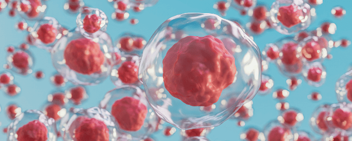
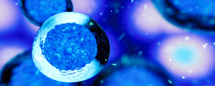
Stem cells are unique cells found in the human body that are the only cells that can differentiate into new cell types. Depending on the need, stem cells can divide and create brain, muscle, and blood cells, as well as various other specialized cells in the body. This ability earned them the label of the “building blocks of cells.”
Stem cells may also help repair damaged tissues, allowing them to play a critical role in managing condition and injury symptoms.
What Are the Types of Stem Cells?
There are two primary forms of stem cells – embryonic and adult.
Embryonic Stem Cells
Embryonic stem cells come from a blastocyst, an early-stage embryo before it implants. In the very early days of forming a human, cells reach a blastocyst stage, which occurs about four to five days after fertilization.
Embryonic stem cells are particularly valuable since they can divide unlimitedly under the right conditions. Since these are the very first cells the body forms to make a human, they can become all types of cells. In addition, they are pluripotent, meaning they can become many types of cells.
The embryonic stem cells used in research now come from unused embryos donated from in-vitro procedures.
Adult Stem Cells
Adult stem cells have two main types. One type of adult stem cell comes from tissue inside the body, such as bone marrow, fat tissue, or skin tissue. However, the number of stem cells in these tissues is limited, and they can only differentiate into specific types of cells, making them multipotent, not pluripotent.
Scientists have discovered ways to change adult stem cells in a lab to make them pluripotent, like embryonic stem cells. These are induced pluripotent stem cells, and they still come from adult tissue.
Stem Cell Therapy
Currently, physicians and scientists may use hematopoietic stem cells (HSCs) or mesenchymal stem cells (MSCs) to help manage conditions. HSCs are adult cells found in the bone marrow. Recently, researchers have found strong evidence that HSCs are pluripotent and may serve as the source of most cells in the body. MSCs are a multipotent cell that can differentiate into different tissues in the body and are found in adipose (fat), umbilical cord, and bone marrow tissues.
While the early years of stem cell research focused on embryonic stem cells, researchers are now uncovering the capability of adult stem cells and discovering new and exciting ways to help patients with managing their conditions and injuries with these unique cells. To learn more about the services offered at Stemedix, contact us today!

by admin | Aug 5, 2022 | Spinal Cord Injury, Mesenchymal Stem Cells, Stem Cell Research, Stem Cell Therapy
Spinal cord injury (SCI) continues to be a significant cause of disability. In fact, it is estimated that annual SCIs account for nearly 18,000 injuries in the United States and between 250,000 and 500,000 injuries worldwide[1]. While the main cause of SCIs in the United States continues to be motor vehicle accidents, other contributors include falls, recreational accidents, and complications from medical procedures.
In their attempt to minimize damage after SCI, researchers have proposed several treatment options. This review conducted by Zoehler and Rebellato identifies cell therapy, and specifically treatment with mesenchymal stem cells (MSCs), as the primary form of neuroregenerative treatment for SCIs.
Research has shown that mammals are unable to regenerate nervous cell tissue in an area damaged as a result of a SCI, which means currently they will be subject to permanent disability after suffering such an injury.
Current treatments for SCIs have proven unable to repair the damage, rather they are used to relieve SCI-associated symptoms, including pressure and scarring, while also attempting to reduce hypoxia resulting from edema and hemorrhaging. One such treatment, spinal compression surgery, has shown to be successful at achieving these outcomes with results being much better if the surgery is completed within 24-hours of the SCI.
Another treatment currently used after SCI is methylprednisolone sodium succinate (MPSS) administered intravenously. In addition to inhibiting lipid peroxidation, MPSS inhibits post-traumatic spinal cord ischemia, supports aerobic energy metabolism, and attenuates neurofilament loss. However, because this treatment is associated with gastrointestinal bleeding and infection, it is recommended to be used with caution.
While not yet fully understood, cell therapy – and specifically therapy using MSCs – has presented promising findings related to regenerating tissue after a SCI. It is widely believed that MSCs effectiveness is related to their ability to secrete different factors and biomolecules.
MSCs also reduce inflammation, which is important in this application because inflammation is known to be a secondary event after sustaining initial SCI.
The authors point out that a better understanding of the specific mechanisms related to the regenerative effects of MSCs used when treating SCI is required in order to develop future MSC-based treatments designed to address SCI in humans. Currently, despite the recent increased focus on the use of cell therapy to treat SCI and central nervous system trauma, there is no consensus on a number of essential topics, including cell type, source, number of cells infusion pathways, and number of infusions to achieve this goal.
Zoehler and Rebellato also point out that it’s important to better understand how the reorganization of injured neural tissues associated with MSCS is related to the restoration of neural function.
Numerous animal model and human clinical trials have confirmed the regenerative and neuroprotective potential of MSCs without adverse effects during or after infusion. The authors close this review by highlighting that MSCs continue to demonstrate potential as an alternative for SCI therapy, primarily because the therapy is not limited by the time of injury and has shown measurable improvements in patients with complete and incomplete SCI.
Source: Fracaro L, Zoehler B, Rebelatto CLK. Mesenchymal stromal cells as a choice for spinal cord injury treatment. Neuroimmunol Neuroinflammation 2020;7:1-12. http://dx.doi.org/10.20517/2347-8659.2019.009
[1] “Spinal cord injury – WHO | World Health Organization.” 19 Nov. 2013, https://www.who.int/news-room/fact-sheets/detail/spinal-cord-injury.

 St. Petersburg, Florida
