by admin | Mar 4, 2025 | Exosomes, Spinal Cord Injury, Stem Cell Research, Stem Cell Therapy
A spinal cord injury (SCI) is a serious condition that affects the central nervous system, leading to loss of movement, sensation, and bodily functions below the site of the injury. SCI is not only life-changing for those affected but also presents a significant burden on healthcare systems worldwide. Each year, thousands of people experience SCI due to accidents, falls, or medical conditions, and unfortunately, there is currently no way to fully restore lost function.
After an SCI occurs, the damage progresses in two stages: primary and secondary injury. The primary injury happens immediately upon impact, causing direct harm to the spinal cord. This is followed by secondary injury, a complex process where inflammation, cell death, and scar formation make it even more difficult for the spinal cord to heal.
In this review, Yu et al. review how exosomes are prepared, their functions, administration routes, and their role in repairing SCI, including their effectiveness alone and in combination with other treatments.
Understanding Exosomes: Functions, Benefits, and Applications
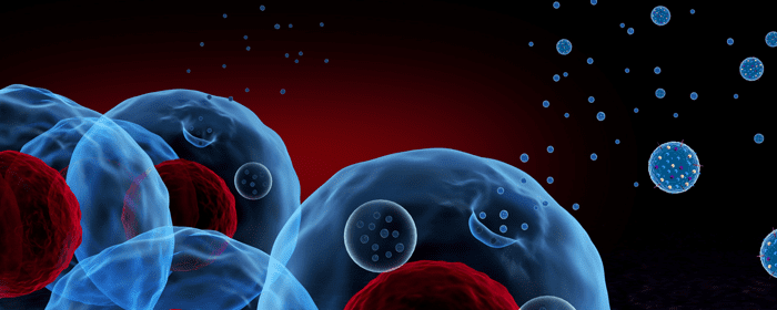
Exosomes are tiny particles that cells release into their surroundings. These microscopic vesicles, which range in size from 30 to 150 nanometers, help cells communicate by carrying proteins, genetic material, and other molecules from one cell to another. Exosomes play a key role in many biological processes, including immune responses, tissue repair, and even disease progression.
According to the authors, scientists have recently begun exploring the potential of exosomes in medicine, particularly for treating spinal cord injuries. Since exosomes are naturally produced by cells and can travel throughout the body, they have the potential to serve as powerful tools for healing damaged tissues, reducing inflammation, and encouraging nerve regeneration.
How Exosomes Can Help Repair SCI
Promoting Nerve Regeneration
One of the most notable challenges in SCI recovery is nerve regeneration. Nerve cells, or neurons, do not repair themselves easily after damage. However, research has shown that exosomes may help stimulate this process. Certain types of exosomes have been found to contain molecules that encourage nerve cell growth and survival. By delivering these molecules to injured areas, exosomes may promote the repair of damaged nerves and improve functional recovery.
Reducing Inflammation
Inflammation is a major contributor to secondary injury after SCI. When the spinal cord is damaged, immune cells rush to the site, releasing chemicals that cause swelling and further harm to nerve cells. Exosomes have been shown to help regulate the immune response by reducing inflammation and preventing excessive damage. By controlling the body’s inflammatory reaction, exosomes may create a more favorable environment for healing.
Protecting Against Cell Death
After SCI, many nerve cells die due to stress and lack of oxygen. Exosomes may offer protection by delivering molecules that help cells survive. Some exosomes have been found to block pathways that lead to cell death, allowing more neurons to stay alive and functional. This protective effect could be crucial in limiting the long-term effects of SCI.
Encouraging Blood Vessel Growth
Blood flow is essential for delivering oxygen and nutrients to the spinal cord. After an SCI, blood vessels in the area may be damaged, further reducing the chances of recovery. Exosomes have been found to support the growth of new blood vessels, improving circulation to injured areas. This process, known as angiogenesis, can help supply the spinal cord with the nutrients it needs to repair itself.
Combating Oxidative Stress
Oxidative stress is another factor that worsens spinal cord injuries. It occurs when harmful molecules called free radicals accumulate and damage cells. Exosomes contain antioxidants that can neutralize these harmful molecules, protecting nerve cells from additional damage. By reducing oxidative stress, exosomes may help preserve spinal cord function and promote healing.
Using Exosomes for SCI Treatment
Direct Injection
One way to use exosomes for SCI treatment is by injecting them directly into the injured area. This method allows exosomes to reach damaged nerve cells quickly and begin their repair work. However, one challenge with this approach is that exosomes may not stay in place long enough to have a lasting effect. Scientists are working on ways to improve the stability and effectiveness of direct injections.
Intravenous Delivery
Another method is intravenous (IV) delivery, where exosomes are injected into the bloodstream. This allows them to travel throughout the body and potentially reach the spinal cord. While IV delivery is less invasive than direct injection, some exosomes may be filtered out by organs like the liver before they reach the injury site. Researchers are exploring ways to improve targeting so that more exosomes reach the spinal cord.
Exosomes Combined with Biomaterials
Scientists are also investigating the use of biomaterials, such as hydrogels, to help exosomes stay at the injury site longer. Hydrogels are soft, water-based materials that can hold exosomes in place, slowly releasing them over time. This controlled release may enhance the effectiveness of exosome therapy and provide a more sustained healing effect.
The Future of Exosome Therapy for Spinal Cord Injury
According to Yu et al. emerging research suggests that exosomes could play a crucial role in promoting healing and improving recovery.
While there are still many questions to answer and challenges to overcome, the authors conclude the potential of exosomes in medicine is undeniable. With continued research and development, exosome therapy could one day provide a groundbreaking solution for spinal cord injury patients, helping them regain function and improve their quality of life.
Source: Yu, T., Yang, LL., Zhou, Y. et al. Exosome-mediated repair of spinal cord injury: a promising therapeutic strategy. Stem Cell Res Ther 15, 6 (2024). https://doi.org/10.1186/s13287-023-03614-y

by admin | Feb 25, 2025 | Back Pain, Exosomes, Regenerative Medicine, Stem Cell Research, Stem Cell Therapy
Low back pain is a widespread issue that affects millions of people worldwide, significantly impacting their daily lives and placing a substantial financial strain on the healthcare system. Existing treatment options for low back pain often provide only temporary relief and come with various limitations. With the increasing interest in regenerative medicine, newer treatments like orthobiologics, including extracellular vesicles or exosomes derived from mesenchymal stem cells, are being explored as potential alternatives for managing musculoskeletal conditions such as low back pain.
As part of this review, Gupta examines the outcomes of clinical studies using extracellular vesicles or exosomes for treating low back pain.
Understanding Low Back Pain
Low back pain is one of the leading causes of disability across the globe, affecting hundreds of millions of people. The condition is expected to increase in prevalence, with estimates suggesting that 843 million people will be affected by 2050. The lifetime risk of experiencing low back pain ranges between 65% and 85%, contributing to over $50 billion in healthcare costs each year.
Several factors can contribute to low back pain, including:
- Lumbar facet joint issues: These joints in the spine can degenerate due to aging, inflammation, or trauma, leading to chronic pain conditions.
- Disc herniation: This occurs when the spinal disc bulges into the spinal canal, compressing nerve roots and causing symptoms such as lumbar radiculopathy (pain radiating from the lower back to the legs).
Traditional Treatments for Low Back Pain
Common treatments for low back pain include physical therapy, chiropractic care, acupuncture, pain-relieving medications (such as narcotics and anti-inflammatory drugs), and minimally invasive procedures like nerve blocks and radiofrequency ablation. Despite their widespread use, these approaches often have limited effectiveness in providing long-term pain relief and may carry side effects. For example, steroid injections—one of the most commonly used interventions—often do not offer significant benefits compared to a placebo.
Emerging Treatments: The Role of Exosomes and Extracellular Vesicles
Recent research has focused on cellular therapies using mesenchymal stem cells (MSCs) due to their ability to regenerate damaged tissues. Extracellular vesicles (EVs), including exosomes, are small particles released by MSCs that play a key role in their therapeutic effects. These vesicles are known to:
- Reduce inflammation: EVs can decrease inflammation by promoting the healing type of immune cells (M2 macrophages).
- Promote tissue repair: They aid in the healing process and have lower risk of immune rejection than the cells themselves.
EVs may overcome some of the limitations of stem cell therapies, such as poor survival and retention at the treatment site, by delivering therapeutic molecules directly to the affected areas. This makes them a promising candidate for treating conditions like low back pain.
Review of Clinical Studies Using Exosomes for Low Back Pain
This review, Gupta looked at studies published up to March 2024 to assess the use of extracellular vesicles and exosomes in treating low back pain. Several databases were searched for relevant studies, including Scopus, PubMed, and Web of Science. The inclusion criteria focused on clinical trials that involved the use of exosomes for low back pain, while studies that did not explicitly use exosomes or were unrelated to low back pain were excluded.
Only two studies met the criteria:
- Study by Phillips et al.: This research involved administering exosomes derived from bone marrow stem cells to patients experiencing lumbar and cervical radiculopathy (nerve pain in the back and neck). The treatment was found to be safe and showed a reduction in pain and improvement in function at a one-month follow-up.
- Study by Wilson et al.: In this study, exosomes were injected into the facet joint space of patients with lumbar facet joint pain. The results indicated that the treatment was safe and led to significant improvements in pain relief and function at a three-month follow-up.
These findings align with other literature supporting the potential benefits of using stem cell-based therapies for managing low back pain. The use of exosomes may provide an effective alternative by retaining the regenerative properties of MSCs while avoiding some of the challenges associated with using live cells.
Exosomes: A Promising Treatment for Low Back Pain
Gupta’s review of current studies suggests that exosomes or extracellular vesicles could offer a safe and potentially effective treatment for low back pain. By targeting inflammation and promoting tissue healing, exosomes may provide a novel approach to managing a condition that affects millions of people. However, further high-quality research is necessary to confirm their long-term safety and effectiveness and to understand how they compare to existing treatments.
Source: Gupta A. Exosomes for the Management of Low Back Pain: A Review of Current Clinical Evidence. Cureus. 2024 Apr 3;16(4):e57539. doi: 10.7759/cureus.57539. PMID: 38707134; PMCID: PMC11068073.

by admin | Oct 25, 2024 | Exosomes, Regenerative Medicine, Stem Cell Research, Stem Cell Therapy
Respiratory diseases are a major global health concern, responsible for millions of deaths each year. Conditions like chronic obstructive pulmonary disease (COPD), acute respiratory distress syndrome (ARDS), pulmonary fibrosis, and pneumonia claim many lives annually, and despite advancements in medical research, there is still no cure for many of these diseases. Current treatments typically focus on managing symptoms and slowing disease progression, but there is growing interest in stem cell (SC) therapy as a potential game-changer for treating lung diseases.
Stem cell therapy is a type of regenerative medicine where stem cells, which have the ability to regenerate or repair damaged tissues, are introduced into the body. There are four main sources of stem cells: embryonic tissues, fetal tissues, adult tissues (like mesenchymal stem cells or MSCs), and genetically manipulated somatic cells, known as induced pluripotent stem cells (iPSCs). Numerous studies have shown that stem cell therapies could be safe and effective for a variety of lung diseases, including COPD, ARDS, and pulmonary fibrosis.
Researchers are increasingly focusing on a cell-free approach that uses stem cell-derived exosomes (SC-Exos). Exosomes are small particles that stem cells release into the body to help with intercellular communication. These exosomes contain beneficial properties of stem cells, including immunomodulatory, anti-inflammatory, and antifibrotic effects.
SC-Exos offer several advantages over stem cell therapy. They have a unique ability to inherit the molecular patterns of their parent stem cells, which means they can potentially mimic the therapeutic effects of stem cells.
Several studies have demonstrated that SC-Exos may be particularly useful for treating respiratory diseases. For example, preclinical and clinical studies have explored the potential of SC-Exos for treating COVID-19, an illness that severely impacts the respiratory system. SC-Exos have been shown to help reduce the severity of complications, such as pneumonia and ARDS, by modulating the immune system and reducing inflammation. In one clinical trial, the use of SC-Exos from bone marrow-derived mesenchymal stem cells (BMSCs) improved survival rates, oxygenation, and immune system regulation in patients.
To deliver SC-Exos effectively, researchers have explored various methods, including intratracheal instillation (direct delivery into the lungs via a tube) and inhalation through nebulizers. Inhalation has shown particular promise, as it allows the exosomes to directly reach the affected lung tissues. In one study involving a mouse model of lung injury caused by the bacterium Pseudomonas aeruginosa, inhaling MSC-derived exosomes significantly improved survival rates. Clinical trials are currently underway to determine if similar results can be achieved in humans.
While many studies attribute the benefits of SC-Exos to their RNA content, it is likely that other components of exosomes also play important roles in their therapeutic effects. Further research is needed to better understand these mechanisms and to optimize the use of exosomes in clinical practice.
Another area of research is focused on developing synthetic or “exosome-mimic” particles that could replicate the therapeutic effects of natural exosomes. These particles could be designed to contain the key bioactive molecules responsible for the beneficial effects of SC-Exos, while being easier and cheaper to produce. However, creating these synthetic particles will require extensive research to ensure they are safe and effective.
Looking ahead, researchers are optimistic about the future of SC-Exos as a potential treatment for respiratory diseases. As our understanding of exosome biology continues to grow, it is likely that we will see more clinical trials and eventually the development of new therapies based on exosome technology. In particular, the use of aerosolized SC-Exos delivered via inhalation holds great promise for treating lung diseases, as it allows the exosomes to directly target damaged tissues in the lungs.
Azhdari et. al conclude that SC-Exos represent an exciting new frontier in the treatment of respiratory diseases. With further research and development, they could offer a powerful new tool for managing and potentially curing conditions like COPD, ARDS, and pulmonary fibrosis, providing hope to millions of patients around the world.
Source: Azhdari MH, Goodarzi N, Doroudian M, MacLoughlin R. Molecular Insight into the Therapeutic Effects of Stem Cell-Derived Exosomes in Respiratory Diseases and the Potential for Pulmonary Delivery. International Journal of Molecular Sciences. 2022; 23(11):6273. https://doi.org/10.3390/ijms23116273

by admin | Feb 15, 2024 | Osteoarthritis, Exosomes, Extracellular Vesicles, Mesenchymal Stem Cells, Stem Cell Research, Stem Cell Therapy
Osteoarthritis (OA) is the most common form of arthritis and is estimated to affect nearly 365 million people worldwide. Characterized as an inflammatory disease, OA slowly progresses over time and results in the gradual loss of the protective cartilage found on the ends of the bones.
While the specific cause of OA has yet to be determined, a growing body of evidence suggests the chondrocyte inflammatory response resulting from elevated levels of pro-inflammatory cytokines is a critical factor in the development and progression of OA.
Recent evidence also suggests that mesenchymal stem cell-derived exomes (MSCs-Exos) exhibit beneficial anti-inflammatory responses in several inflammatory diseases, including OA.
In this study, Wang et al. explore the role of human umbilical cord-derived MSCs-Exos (hUC-MSCs-Exos) in treating the inflammation of chondrocytes and its related mechanisms.
As part of this study, the authors report that supplementing the observed chondrocyte inflammation models with hUC-MSCs-Exos demonstrated the ability to reduce the inflammation of chondrocytes caused by the inflammatory factor IL-1β.
Additionally, activation and polarization of synovial macrophages to M1 phenotypes also contribute to the progression of OS. As part of this study, Wang et al. report that hUC-MSC-Exos demonstrated a protective effect against M1 macrophage-induced chondrocyte damage and cell death.
Wang et al. indicate that the results of this study confirm the anti-inflammatory effects of hUC-MSCs-Exos in the human articular chondrocytes inflammation model. The authors also conclude that hUC-MSCs-Exos may be used as a potential cell-free treatment for chondrocyte inflammation in OA.
Source: Wang S, Jiang W, Lv S, et al. Human umbilical cord mesenchymal stem cells-derived exosomes exert anti-inflammatory effects on osteoarthritis chondrocytes. Aging (Albany NY). 2023;15(18):9544-9560. doi:10.18632/aging.205034

by admin | Jan 17, 2024 | Exosomes, Glaucoma, Regenerative Medicine, Stem Cell Research, Stem Cell Therapy
According to the World Health Organization, at least 2.2 billion people worldwide have near or distant vision impairment. In at least 1 billion of these cases, vision impairment could have been prevented or has yet to be addressed. Glaucoma, corneal disease, and retinal disorders are among the leading contributors to these vision impairments.
Since the pathogenesis of these eye diseases is not fully understood, fully effective treatments have yet to be developed. Considering this, Li et al. reviewed recent research to examine the effectiveness of exosomes in various diseases in vivo, which provides the potential for a new option for the treatment of eye diseases.
Exosomes are extracellular small vesicles that are formed by the regulation of endocytosis, fusion, and efflux and contain a variety of biologically active substances, including proteins, miRNAs, IncRNAs, and lipids.
Exosomes are found in all biological fluids and have roles that vary depending on their origin. These roles include cell-to-cell communication, waste transfer, and regulation of the immune system in vivo. Additionally, when serving as a carrier, exosomes are involved in many pathological processes such as nerve repair, vascular regeneration, immune response, and fibrosis formation.
Examining the various roles exosomes play within the body, the authors of this review consider their role in the treatment of serious ocular diseases, including glaucoma, diabetic retinopathy, and keratitis. Li et al. point to studies demonstrating exosomes’ ability to promote the repair of injured nerves, inhibit fibrosis, modulate immune function, and promote angiogenesis as evidence of the important role they have in treating ocular disease.
Specifically, exosomes contain a large number of immunosuppressive molecules that inhibit lymphocyte proliferation and effectively increase ocular immune tolerance to prevent ocular autoimmune disease.
Exosomes can also transfer protein and RNA to receptor cells and can accelerate wound healing of corneal epithelial cells, providing a new approach for treating large corneal lesions.
Studies have also demonstrated a link between exosomes and age-related macular degeneration (AMD) with specific exosomes considered to have neuroprotective effects that are closely related to the pathological progression of AMD.
The authors conclude that exosomes are able to be used as therapeutic carriers to participate in processes such as immune response, angiogenesis, and nerve repair in ocular-related diseases. While research into this is still emerging, the presence and accessibility of exosomes will become a potential way to diagnose and treat ocular diseases.
Source: “Progress in exosomes and their potential use in ocular diseases.” 18 Sep. 2020, https://www.ncbi.nlm.nih.gov/pmc/articles/PMC7459212/.

by admin | Jan 11, 2024 | Exosomes, Stem Cell Research, Stem Cell Therapy
Repairing the structure and functionality associated with subcutaneous cartilage defects continues to be a challenge in the fields of plastic and reconstructive surgery. While current methods, including autologous chondrocyte implantation and matrix-assisted chondrocyte implantation, have been successful in some regard, they continue to present a number of limitations, including donor limitation, donor morbidity, and degradation of the graft tissue.
Recently, cartilage progenitor cell (CPC)-based tissue engineering has drawn attention in the field of cartilage regeneration, primarily for its strong chondrogenic differentiation capacity.
Unfortunately, a general lack of a suitable chondrogenic niche has continued to hinder the clinical application of CPC-regenerated cartilage in the subcutaneous environment.
Considering this, and for the purposes of this study, Chen et al. explored the use of exosomes derived from chondrocytes (CC-Exos) as a way to provide the CPC constructs with a cartilage signal in subcutaneous environment for efficient ectopic cartilage regeneration.
After 12 weeks of post-surgical injection of CC-Exos, the authors’ animal model demonstrated that the CC-Exos injections effectively increased collagen deposition and minimized vascular ingrowth in engineered constructs, which efficiently and reproducibly developed into cartilage. This study also demonstrated that the CPC constructs supplied with these CC-Exos could also form cartilage-like tissue with minimal hypertrophy in a subcutaneous environment and with no help from any chondrogenic factors.
Additionally, Chen et al.’s study showed that CC-Exos significantly promoted chondrogenesis-related factors at the mRNA and protein levels in CPCs while also limiting angiogenesis typically associated with hypertrophic differentiation and subsequent calcification.
Despite these promising results, Chen et al. point out that the exact components associated with CC-Exos have yet to be determined. Because of this, the authors call for additional studies to determine the specific components of CC-Exos and their underlying mechanisms related to cartilage repair.
Considering the findings of this study, the authors believe that CC-Exos alone could provide a preferable chondrogenic environment, help maintain the stability of cartilage tissue, and serve as a promising therapeutic approach for the treatment of ectopic cartilage defects.
Source: Chen, Y., Xue, K., Zhang, X. et al. Exosomes derived from mature chondrocytes facilitate subcutaneous stable ectopic chondrogenesis of cartilage progenitor cells. Stem Cell Res Ther 9, 318 (2018). https://doi.org/10.1186/s13287-018-1047-2


 St. Petersburg, Florida
