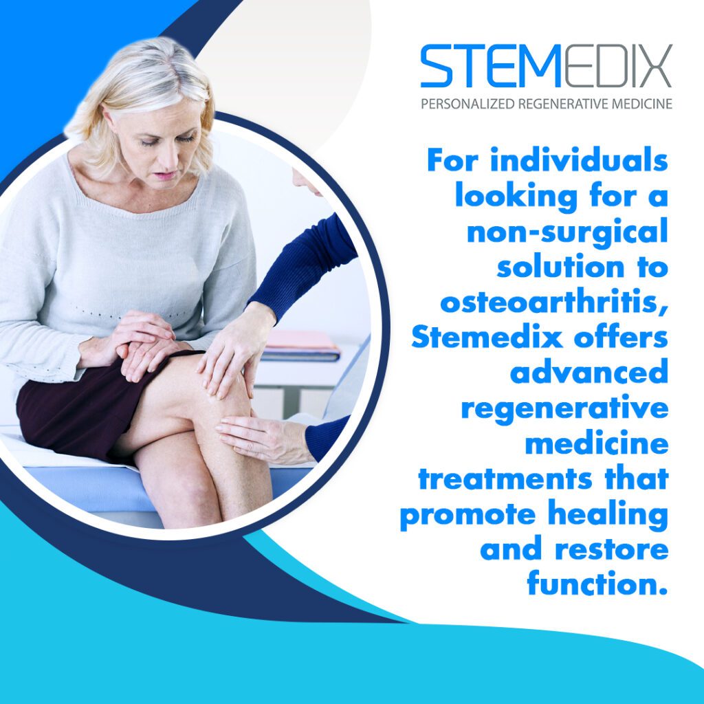
Osteoarthritis can make everyday movements difficult, limiting mobility and causing persistent joint pain. If you have been relying on pain medications or considering surgery, there is another option. At Stemedix, we offer regenerative medicine treatments that address joint damage at the source, helping you regain function without invasive procedures.
Through therapies like stem cell and platelet-rich plasma (PRP) injections, regenerative medicine promotes natural healing by stimulating tissue repair and reducing inflammation. Unlike traditional treatments that only manage symptoms, these therapies work to restore cartilage and improve joint function over time.
For those seeking regenerative medicine in Saint Petersburg, FL, Stemedix offers advanced, full-service, patient-focused care tailored to each individual’s needs. From airport and appointment transportation to providing wheelchairs, walkers, and shower chairs, Stemedix guarantees a comfortable and supported experience throughout your treatment journey. Our team of experts develops personalized treatment plans designed to help you stay active and avoid surgery.
Osteoarthritis and Its Impact on Joint Health
Osteoarthritis (OA) is a progressive joint disease that causes cartilage deterioration, leading to pain, stiffness, and mobility issues. It is the most common type of arthritis, affecting millions of people worldwide. It develops as the protective cartilage that cushions the ends of bones begins to wear down. Over time, this deterioration leads to increased friction between bones, causing pain, stiffness, and inflammation. The condition primarily affects joints that bear the most weight, such as the knees, hips, and spine. However, it can also impact the hands and other areas, making even simple movements uncomfortable.
Although aging plays a role in its progression, osteoarthritis is not just a condition that affects older adults. Joint injuries, repetitive stress, obesity, and genetic factors can contribute to early onset and severity. Many individuals begin to notice symptoms when stiffness becomes more persistent, and activities that were once effortless start to feel more challenging. Without intervention, the condition can continue to worsen, affecting overall mobility and quality of life.
How Osteoarthritis Affects Joint Function and Mobility
Joints rely on smooth cartilage surfaces for pain-free movement. As osteoarthritis progresses, cartilage breaks down, exposing the underlying bone. This increases friction during movement, triggering inflammation and pain. Swelling and stiffness can make it difficult to bend, straighten, or bear weight.
As osteoarthritis advances, bone spurs can form around the joint, restricting motion and adding to discomfort. Fluid accumulation within the joint capsule contributes to swelling, further limiting movement. In severe cases, the cartilage loss becomes so pronounced that bones grind directly against each other, resulting in chronic pain and mobility impairments.
The impact of osteoarthritis extends to daily activities. Tasks like walking, climbing stairs, or standing for long periods become increasingly difficult. Individuals may also experience weakness or instability, increasing the risk of falls or further injury, which can substantially affect independence and overall well-being.
For those struggling with osteoarthritis, finding effective treatment options is essential. At Stemedix, we provide regenerative medicine treatments designed to support natural joint repair, helping individuals regain function and reduce pain without relying solely on medications or surgery.
The Role of Regenerative Medicine in Osteoarthritis Treatment
Regenerative medicine is an approach that utilizes the body’s natural healing mechanisms to repair and restore damaged tissues. This includes stem cell therapy and specialty cells, which can replace and rebuild cartilage, reduce inflammation, and improve joint function, which help regenerate cartilage, reduce inflammation, and improve joint function.
How Stem Cell Therapy Can Aid in Joint Repair
Stem cell therapy introduces specialized cells into the affected joint to help repair and regenerate damaged tissue. These cells are typically sourced either from the patient’s own bone marrow or adipose (fat) tissue, or from carefully screened donor tissue, depending on the treatment approach. Once introduced into the joint, they interact with the surrounding environment to promote healing. Studies suggest that stem cells, especially chondrocytes (Cartilage Cells) and their respective exosomes can contribute to cartilage regeneration, slow the progression of osteoarthritis, and improve overall joint function.
One of the main benefits of stem cell therapy is its ability to address joint degeneration at a cellular level. Unlike pain medications or corticosteroid injections, which only provide temporary symptom relief, stem cells work to support the restoration of tissue over time. Many individuals who undergo stem cell therapy report gradual improvements in pain levels, joint flexibility, and overall mobility. While results vary, research continues to explore how stem cell treatments can enhance long-term joint health.
Platelet-rich plasma (PRP) Therapy: A Complementary Treatment
PRP therapy is another regenerative medicine treatment that can support joint healing. This therapy involves drawing a small sample of the patient’s blood, processing it to concentrate the platelets, and injecting the enriched plasma into the affected joint. Platelets contain growth factors that stimulate tissue repair, reduce inflammation, and enhance the body’s natural healing processes.
For osteoarthritis patients, PRP therapy can help manage pain and improve function by promoting cartilage repair. When used in combination with stem cell therapy, PRP may enhance the effects of treatment by creating an environment that supports regeneration. Some individuals experience noticeable improvements in joint comfort and mobility after a series of PRP injections, making it a valuable option for those seeking alternatives to surgery.
At Stemedix, we provide regenerative medicine treatments designed to support joint repair and improve quality of life. By offering stem cell therapy and PRP therapy, we help individuals take a proactive approach to osteoarthritis management, reducing pain and enhancing mobility without the need for invasive procedures.
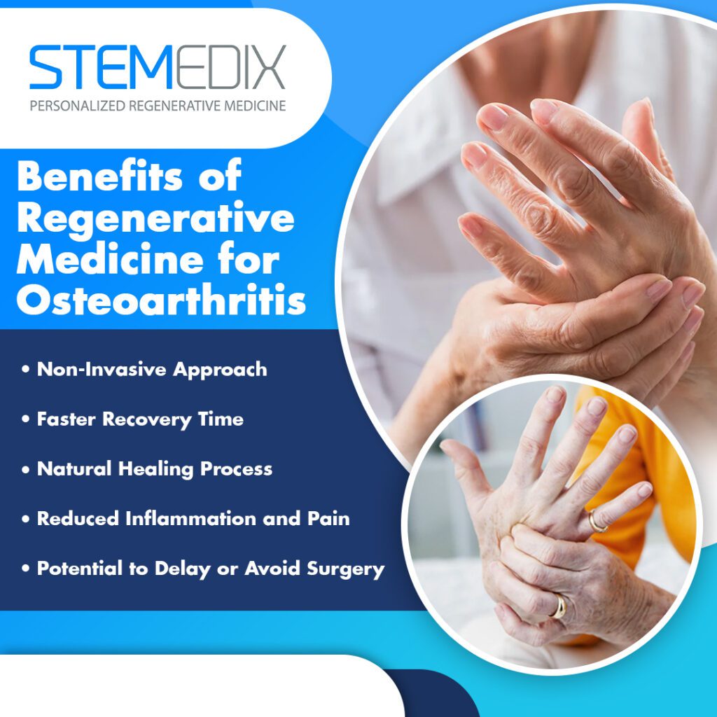
Benefits of Regenerative Medicine for Osteoarthritis
Regenerative medicine treatments provide several benefits for osteoarthritis patients looking for a non-surgical solution:
Non-Surgical Treatment with Minimal Recovery Time
Regenerative medicine treatments offer a non-invasive approach to managing osteoarthritis. Unlike joint replacement surgery, which requires extensive incisions, hospital stays, and long recovery periods, regenerative therapies involve targeted injections. This means patients experience minimal discomfort during the procedure and can return to their normal activities much sooner. Most individuals can resume daily routines within days, making it a practical option for those who want to avoid the risks and downtime associated with surgery.
Because these treatments do not require general anesthesia or large incisions, the risk of complications is lower compared to surgical interventions. Many patients choose regenerative medicine to maintain an active lifestyle while managing osteoarthritis symptoms effectively.
Promoting Natural Healing and Tissue Regeneration
Traditional osteoarthritis treatments, such as pain medications and steroid injections, primarily focus on symptom relief. While they may reduce discomfort temporarily, they do not contribute to long-term joint health. Regenerative medicine takes a different approach by supporting the body’s natural ability to heal itself.
Stem cell therapy and PRP therapy introduce healing elements directly into the affected joint, encouraging the repair of damaged cartilage and tissues. Stem cells can develop into various types of cells needed for joint repair, while PRP provides growth factors that stimulate tissue regeneration. Over time, these therapies may help slow osteoarthritis progression, preserving joint function and mobility for longer.
Reducing Pain and Inflammation
Inflammation is one of the primary drivers of osteoarthritis pain. As the cartilage wears down and bones begin to rub together, the body responds with increased inflammation, leading to swelling, stiffness, and discomfort. Regenerative medicine treatments target this underlying inflammation rather than simply masking pain.
By introducing stem cells and PRP into the affected joint, these treatments help regulate the inflammatory response. This can lead to sustained pain relief and improved joint function over time. Many individuals report noticeable reductions in pain and stiffness, allowing them to engage in physical activities with greater ease.
At Stemedix, we specialize in regenerative medicine treatments that help individuals manage osteoarthritis without relying on invasive procedures. By offering innovative stem cell therapies that promote healing, reduce inflammation, and restore function, we provide a path to long-term joint health and improved mobility.
Is Regenerative Medicine the Right Treatment for You?
Regenerative medicine is an effective treatment option for individuals with mild to moderate osteoarthritis who are seeking a non-surgical alternative to managing their condition. If you have joint pain, stiffness, and decreased mobility that have not responded well to traditional treatments, regenerative therapies may be an excellent choice.
This approach is especially suited for people who wish to avoid the risks, recovery time, and complications associated with joint replacement surgery. If you are looking to regain function, reduce pain, and maintain mobility without the need for invasive procedures, regenerative medicine may be the solution for you.
What to Expect During the Treatment Process
The process for regenerative medicine treatments typically involves injecting stem cells, specialty cells, or even platelet-rich plasma into the affected joint. The procedure is minimally invasive, usually performed in an outpatient setting, and involves very little discomfort.
Before the injection, your healthcare provider will make sure that the area is numbed to minimize any discomfort during the procedure. The stem cells, or PRP, are then injected directly into the affected joint. While the treatment itself is relatively quick, it may take several weeks for noticeable improvements to appear. Over the following months, the benefits of the therapy continue to unfold, helping to promote tissue regeneration, reduce inflammation, and alleviate pain.
At Stemedix, we specialize in providing personalized regenerative medicine treatments tailored to your unique needs. If you’re considering this option for osteoarthritis, our experienced team can help guide you through the process, ensuring that you receive the best care and support throughout your treatment journey.
Regenerative Medicine vs. Traditional Joint Replacement Surgery
Joint replacement surgery becomes necessary when osteoarthritis has progressed to a point where the damage to the joint is severe and non-surgical treatments no longer provide relief. In these cases, the cartilage has deteriorated, causing bones to rub against each other, resulting in debilitating pain and limited movement. When conservative treatments like medication, physical therapy, and injections fail to alleviate symptoms, joint replacement may be the only remaining option. This procedure involves removing the damaged cartilage and bone and replacing it with an artificial joint.
However, joint replacement surgery comes with risks, including infection, blood clots, and a lengthy recovery period. For many patients, the idea of undergoing surgery and spending months recovering is stressful.
Advantages of Regenerative Medicine Over Surgery
Regenerative medicine offers a compelling alternative to joint replacement by addressing joint deterioration early in the process. By harnessing the body’s natural healing abilities, regenerative treatments like stem cell therapy and PRP (Platelet-Rich Plasma) therapy target the root causes of joint pain and inflammation.
One of the most notable benefits of regenerative medicine is the shorter recovery time compared to surgery. Since regenerative treatments involve injections rather than incisions, patients typically experience less pain and minimal downtime and can return to their daily activities much faster. Additionally, regenerative medicine carries fewer risks, as it’s a non-invasive procedure that doesn’t require general anesthesia or long hospital stays.
Another significant advantage is that regenerative treatments may help delay or even prevent the need for joint replacement altogether. By stimulating tissue regeneration, regenerative medicine can slow the progression of osteoarthritis and preserve joint function.
Cost Considerations: Regenerative Medicine vs. Joint Replacement
While joint replacement surgery can cost tens of thousands of dollars, regenerative medicine treatments tend to be more affordable. Surgery often requires extensive post-operative care, including physical therapy and follow-up visits, which can add up in costs. Additionally, joint replacement surgery typically involves a long recovery period, where additional medical expenses may arise.
On the other hand, regenerative medicine offers a cost-effective alternative. Since the procedures are minimally invasive, they generally don’t require the same level of post-treatment care and rehabilitation. By avoiding the need for surgery, patients can save money in the long term, while also potentially experiencing better outcomes without the extensive recovery times.
At Stemedix, we provide advanced regenerative medicine treatments tailored to your needs, offering an affordable, non-surgical alternative that may help you avoid joint replacement surgery. Our team of experienced providers is here to guide you through your treatment options and help you make the best decision for your joint health.
Stemedix: Your Partner in Regenerative Medicine for Osteoarthritis
At Stemedix, we specialize in offering advanced regenerative medicine in Saint Petersburg, FL, designed to provide relief from osteoarthritis and restore joint function. Our clinic is known for its commitment to providing the highest level of care, using innovative treatments like stem cell therapy, specialty cells, and PRP (plat-r-p) therapy. These treatments are tailored to help manage pain, reduce inflammation, and regenerate damaged tissues, ultimately improving mobility and quality of life.
We understand that each patient’s needs are unique, which is why we focus on personalized care and treatment options. Whether you’re looking to delay surgery or improve your joint function, Stemedix is dedicated to helping you achieve your goals with effective, non-surgical alternatives.
How Stemedix Tailors Treatments for Osteoarthritis Patients
At Stemedix, we take a personalized approach to every case of osteoarthritis. Our medical team works closely with each patient to design a customized treatment plan that suits their specific condition. We don’t believe in a generalized approach—our regenerative medicine treatments are based on evidence-based practices that are aimed at providing the most effective results for your joint health.
Whether it’s stem cell therapy to regenerate damaged cartilage or PRP therapy to reduce inflammation and promote healing, we combine these treatments to maximize joint restoration and pain relief. Our goal is to enhance your mobility and quality of life while avoiding the need for invasive surgeries.
Patient-Centered Care at Stemedix
At Stemedix, based in Saint Petersburg, FL, patient care is at the heart of everything we do. From the moment you walk through our doors, you’ll experience a welcoming and supportive environment. We understand that going through osteoarthritis treatment options can be overwhelming, which is why we assign dedicated care coordinators to guide you every step of the way.
Our care coordinators make sure that you have all the information you need to make well informed decisions about your treatment. They work closely with you to schedule appointments, answer questions, and monitor your progress throughout the healing process. We prioritize your comfort, convenience, and well-being, ensuring that your experience is smooth and stress-free, from consultation to recovery.
Explore Regenerative Medicine as a Viable Osteoarthritis Treatment Option at Stemedix
Regenerative medicine offers a valuable treatment option for individuals experiencing osteoarthritis in Saint Petersburg, FL. Unlike conventional methods that focus solely on symptom management, regenerative therapies such as stem cell therapy and PRP (Platelet-Rich Plasma) therapy work to restore joint health by promoting natural healing. These treatments support tissue repair, reduce inflammation, and encourage cartilage regeneration, which can enhance mobility and long-term joint function.
Stemedix specializes in regenerative medicine in Saint Petersburg, FL, providing a non-surgical alternative for those seeking relief from osteoarthritis. By leveraging innovative therapies, Stemedix helps patients regain mobility and improve their quality of life without the risks and extended recovery associated with surgery.
If you are looking for an advanced, non-invasive approach to managing osteoarthritis, Stemedix is here to help. Contact us today at (727) 456-8968 or email yourjourney@stemedix.com to learn more about how regenerative medicine can support your joint health.
Tissue engineering is an emerging field within regenerative medicine that seeks to repair or regenerate damaged tissues using principles from biology, engineering, and materials science. Stemedix, a prominent provider of regenerative medicine in Saint Petersburg, Florida, incorporates these advancements into personalized treatments designed to enhance patients’ quality of life.
Tissue engineering relies on key components such as biomaterials, cellular therapies (including stem cells), and growth factors to develop treatments for a variety of conditions, including orthopedic injuries and neurodegenerative disorders. This specialized field of medicine enhances the body’s natural healing processes, offering tailored solutions based on individual patient needs. In this article, we will explore the critical role of tissue engineering in regenerative medicine, its current applications, and how Stemedix is bringing this innovative science to life for patients in Saint Petersburg and beyond.
Understanding Tissue Engineering in Regenerative Medicine
What Is Tissue Engineering?
Tissue engineering is a vital aspect of regenerative medicine, focused on creating functional tissues to repair or replace damaged biological structures. This interdisciplinary field merges biology, engineering, and medicine to create systems that support tissue regeneration and repair within the body. Central to this process are key components: cells, scaffolds, and growth factors, working in unison to support and enhance the body’s natural healing capabilities.
Tissue engineering aims to create tissues that closely replicate the structure and function of natural tissues, thereby supporting the body’s ability to heal itself. Engineered tissues are created using stem cells, various cell types, biocompatible scaffolds, and signaling molecules that promote cell growth, differentiation, and tissue regeneration.
From its initial focus on skin and cartilage repair, tissue engineering has evolved to address complex tissues like bone, nerve, and heart structures, representing significant advancements in regenerative medicine’s potential to improve lives.
How Tissue Engineering Supports Regenerative Medicine
Tissue engineering is central to regenerative medicine by enhancing the body’s ability to heal itself. When tissue damage is severe or chronic, the body’s natural healing processes may fall short. Tissue engineering addresses this challenge by providing essential components for the repair, regeneration, or replacement of damaged tissues.
A cornerstone of this approach involves biomaterials, such as scaffolds, which act as frameworks for cellular growth and tissue organization. Scaffolds replicate the body’s extracellular matrix (ECM), providing both structural and biochemical cues to guide cell growth, differentiation, and tissue development.
Tissue engineering has already made significant strides in treating orthopedic conditions, including cartilage and bone repair. It is also being explored for nerve regeneration, including spinal cord injuries. By leveraging patient-specific cells, these therapies are not only personalized but also reduce the risk of rejection, offering a seamless integration into the body for sustainable and effective healing.
At Stemedix, we apply tissue engineering techniques in our regenerative medicine treatments, supporting patients in Saint Petersburg and beyond on their recovery journey. By focusing on personalized care and applying research-driven approaches, we aim to assist patients in improving function and managing pain. Our team is dedicated to offering clear guidance and support throughout the healing process, working with each individual to find the most appropriate path for their unique needs.
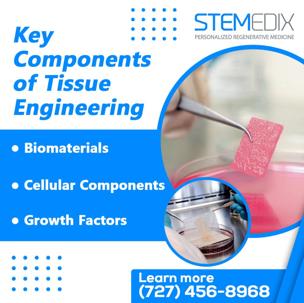
Key Components of Tissue Engineering
Tissue engineering is at the heart of regenerative medicine, combining the expertise of biologists, engineers, and medical professionals to repair and regenerate damaged tissues. To understand how tissue engineering works, it’s important to break down its key components—biomaterials, cellular components, and growth factors. Each plays a crucial role in facilitating the healing process and promoting tissue regeneration.
Biomaterials: The Building Blocks of Tissue Engineering
Biomaterials play a pivotal role in tissue engineering, serving as essential building blocks for supporting the body’s natural healing processes. In regenerative medicine, these materials are utilized to construct scaffolds that provide structural support for tissue regeneration. Acting as a framework, scaffolds enable cells to attach, grow, and differentiate into specific tissue types.
Biomaterials are broadly categorized into natural and synthetic types. Natural biomaterials, such as collagen and hyaluronic acid, are derived from biological sources and integrate seamlessly with the body’s tissues due to their high biocompatibility. Synthetic biomaterials, like engineered polymers, offer customizable properties such as strength, flexibility, and controlled degradation, making them ideal for various tissue regeneration needs.
Beyond structural support, biomaterials replicate the extracellular matrix (ECM), a natural cellular environment crucial for guiding tissue growth and function. By mimicking the ECM, biomaterials ensure proper cell behavior, aiding in the formation of functional, healthy tissues.
Cellular Components: Fueling Regeneration
Cellular components are the driving force behind tissue regeneration, making them indispensable in regenerative medicine. Stem cells, in particular, are vital due to their unique ability to transform into various cell types depending on the specific tissue needing repair. These cells can be sourced from the patient’s own body (autologous stem cells), minimizing immune rejection, or from donor tissues (allogeneic stem cells). Both options are key to tailoring treatments for individual needs.
In addition to stem cells, progenitor cells also play a significant role in tissue engineering. These more specialized cells retain the ability to develop into specific tissue types, such as cartilage, bone, or even neural tissues. Sourcing and cultivating these cells involves advanced techniques. Some are collected directly from the patient, offering a personalized approach, while others are expanded in laboratories to ensure sufficient quantities for treatment. Combined with biomaterials, these cells form scaffolds that support effective tissue regeneration.
Growth Factors: Catalysts for Tissue Development
Growth factors are essential signaling molecules that regulate cellular processes, such as cell growth, differentiation, migration, and tissue remodeling, which are critical for tissue regeneration. These signaling molecules also play a pivotal role in angiogenesis (the formation of new blood vessels) and tissue remodeling, ensuring proper healing and restoration.
In regenerative medicine, growth factors are either directly applied to injured areas or integrated into biomaterials within tissue scaffolds. This approach enhances the body’s natural healing mechanisms, guiding cells to the injury site and promoting accurate tissue formation.
Key examples include vascular endothelial growth factor (VEGF), which supports blood vessel formation; platelet-derived growth factor (PDGF), critical for wound healing; and transforming growth factor-beta (TGF-β), which aids in tissue repair. When combined with stem cells and biomaterials, these growth factors create a synergistic effect, improving the effectiveness of regenerative medicine treatments and fostering comprehensive tissue repair.
Together, these three components—biomaterials, cellular elements, and growth factors—form the foundation of tissue engineering in regenerative medicine. As we continue to develop and refine these technologies, their role in healing and recovery will only expand, providing new hope and opportunities for patients seeking alternative treatment options.
Current Applications in Regenerative Medicine
Regenerative medicine, powered by tissue engineering, is advancing rapidly, offering new methods to repair and regenerate tissues previously considered irreparable. This breakthrough science has numerous applications across various medical fields, including orthopedics, organ and tissue replacement, and neurodegenerative conditions. Below, we explore some of the most significant advancements in regenerative medicine and how they are impacting patient care.
Advancements in Orthopedics
Orthopedic conditions affecting the musculoskeletal system are among the most common areas where regenerative medicine is making significant progress. Cartilage, bones, and tendons are vital structures that can suffer from degeneration or injury, leading to chronic pain and disability. Regenerative medicine treatments, such as stem cell therapy and tissue engineering, are providing innovative solutions to repair and regenerate these tissues.
In orthopedic applications, stem cells are used to promote healing in damaged cartilage and bone, offering the potential for repairing joint injuries, fractures, and degenerative conditions like osteoarthritis. Biomaterials, often used as scaffolds, provide the structural support needed for new tissue to grow, while growth factors stimulate the healing process. For example, stem cells derived from the patient’s own body are applied to injured areas, where they can differentiate into the required cell types, promoting faster and more efficient healing. These advancements in orthopedics help patients recover faster, with fewer complications and less reliance on invasive surgeries.
Innovations in Organ and Tissue Replacement
A promising area of tissue engineering in regenerative medicine is the development of engineered tissues to replace damaged or failing organs. Traditional organ transplantation faces significant challenges, including organ shortages, immune rejection, and long waiting times. Tissue engineering aims to overcome these barriers by developing engineered tissues that can perform the functions of organs like the liver, heart, and kidneys.
For example, regenerative medicine approaches are being tested to create functional liver tissue from stem cells, offering potential treatment options for patients with liver failure. Similarly, engineered cardiac tissue could be used to repair heart damage caused by disease or injury, and advances in kidney regeneration are showing promise for individuals suffering from kidney disease. Through these innovations, the need for organ donations could be reduced, and patients could experience faster recovery times with improved long-term outcomes.
Impact on Neurodegenerative Conditions
Neurodegenerative conditions, such as Alzheimer’s disease, Parkinson’s disease, and spinal cord injuries, present some of the most complex medical challenges. However, tissue engineering and regenerative medicine are offering new hope for patients affected by these conditions. One of the most promising areas of research involves using stem cells and engineered tissues to repair spinal cord injuries and promote brain cell regeneration.
Stem cells have the potential to differentiate into various types of neural cells, which can help repair damaged nerve tissue in the brain and spinal cord. Researchers are also exploring how to stimulate the growth of new neurons in areas of the brain affected by neurodegenerative diseases. Integrating tissue engineering with stem cell therapy holds promise for restoring lost function in the nervous system, offering new treatment options for patients with neurodegenerative diseases or spinal cord injuries.
Regenerative medicine is opening doors to innovative solutions that address some of the most challenging medical conditions. From orthopedic injuries to tissue replacement and neurodegenerative diseases, the potential applications of tissue engineering are vast and continue to expand. At Stemedix, we are proud to be at the forefront of this field, offering advanced treatments that aim to restore health and potentially improve the quality of life for our patients. Through personalized care and the latest advancements in regenerative medicine, we are committed to making a meaningful difference in your health journey.
How Stemedix Advances Regenerative Medicine in Saint Petersburg
As a leader in regenerative medicine in Saint Petersburg, Stemedix is at the forefront of providing innovative therapies that promote healing and improve patients’ quality of life. Our commitment to advancing tissue engineering and regenerative medicine has positioned us as a trusted provider in the region. Through a personalized, patient-centered approach, we utilize pioneering treatments that are scientifically proven to restore function and reduce symptoms of various medical conditions. Let’s explore how Stemedix is advancing regenerative medicine in Saint Petersburg and how these treatments are making a meaningful impact on patients’ lives.
Stemedix’s Approach to Regenerative Therapies
At Stemedix, we are deeply committed to providing regenerative medicine treatments that prioritize both safety and ethical practices. Our treatments are designed not only to meet the highest standards in medical care but also to ensure the best possible outcomes for our patients. Each therapy is carefully selected based on the individual’s unique medical history, goals, and needs.
Our approach is centered around personalized patient care. In Saint Petersburg, patients receive dedicated support from care coordinators, who guide them through every step of the treatment process. From the initial consultation to the post-treatment follow-up, we ensure our patients feel supported and informed throughout their journey. Our team works closely with each patient to develop a tailored treatment plan that incorporates regenerative therapies, such as stem cell treatments, tissue engineering, and growth factor therapy, to address their specific conditions.
By emphasizing personalized care and adhering to ethical practices, Stemedix strives to provide patients in Saint Petersburg with access to high-quality regenerative medicine treatments, supporting their journey toward improved health and wellness.
The Role of Tissue Engineering in Stemedix’s Treatments
Tissue engineering is a cornerstone of the regenerative medicine treatments provided at Stemedix. By leveraging advanced tissue engineering techniques, we offer therapies designed to repair and regenerate damaged tissues, enabling patients to achieve meaningful improvements in their health and well-being.
At Stemedix, our regenerative treatments combine biomaterials, stem cells, and growth factors to facilitate tissue repair. These components work together to restore function in areas affected by injury or disease. For instance, in orthopedic applications, stem cells support cartilage or bone repair, while engineered tissues aid in rebuilding damaged structures.
Similarly, in neurodegenerative conditions, tissue engineering promotes the regeneration of nerve cells in the brain and spinal cord, offering hope for enhanced recovery.
The effect of tissue engineering in our treatments is seen in the positive outcomes experienced by our patients. Many report improved mobility, reduced pain, and an enhanced quality of life. By incorporating these methods, we help individuals in Saint Petersburg reclaim independence and achieve better health.
Why Choose Stemedix for Regenerative Medicine Treatments
When it comes to choosing a provider for regenerative medicine in Saint Petersburg, Stemedix stands out for its unwavering commitment to delivering innovative treatments backed by science. Our patient-centered approach, ethical practices, and expertise in the field ensure that every patient receives the highest level of care. Here’s why Stemedix should be your trusted partner in regenerative medicine.
Ethical and Patient-Centered Care
At Stemedix, we firmly believe that ethical practices and exceptional patient care are the foundation of effective healing. We deeply value the trust our patients place in us, which is why we are committed to transparency, integrity, and compassion in every interaction. Choosing Stemedix means becoming a partner in your healing journey, where your voice matters and your well-being is our priority.
Our dedicated care coordinators are with you every step of the way—from your initial consultation to post-treatment follow-ups—providing personalized support and addressing all your questions. We aim to empower you with clear, accurate information so you can make informed decisions about your health.
Our goal is to create an environment where you feel heard, respected, and cared for, ensuring your experience with regenerative medicine is stress-free and effective. Stemedix is proud to deliver ethical, patient-centered care that prioritizes your unique needs.
Expertise in Regenerative Medicine
Stemedix’s expertise in regenerative medicine is built on years of in-depth research, development, and hands-on experience. Our team of board-certified providers collaborates with patients to create personalized treatment plans tailored to their specific needs. By incorporating the latest advancements in regenerative therapies, including stem cell treatments, tissue engineering, and growth factor therapy, we ensure that our solutions are effective for a wide range of medical conditions.
With a well-established presence in regenerative medicine, Stemedix has earned a reputation for excellence in Saint Petersburg and beyond. We are committed not only to providing high-quality treatments but also to continuously advancing our knowledge. Our ongoing research and partnerships with top biomedical manufacturers allow us to remain at the cutting edge of regenerative medicine, ensuring our patients receive the most effective therapies available.
Whether addressing orthopedic conditions, neurodegenerative diseases, autoimmune disorders, or wellness concerns, Stemedix offers unmatched expertise. Choosing Stemedix means selecting a provider dedicated to ethical practices, personalized care, and proven results. We are here to help guide you toward optimal health and improved quality of life.
Shaping the Future of Healing with Stemedix
Regenerative medicine is transforming healthcare by offering innovative treatments that harness the body’s natural ability to heal. At Stemedix, we are leading the way in providing cutting-edge therapies that not only address the root causes of chronic conditions but also promote faster recovery, improved healing, and an enhanced quality of life.
Our team of experts is dedicated to delivering personalized care, advanced technologies, and research-backed treatments tailored to your unique needs. Whether you are dealing with orthopedic pain, neurodegenerative diseases, or seeking overall wellness support, Stemedix is here to help you navigate your path to better health. Take control of your healing journey today. Reach out to our team for a consultation and discover how our regenerative medicine treatments can improve your well-being. Contact Stemedix at (727) 456-8968 or yourjourney@stemedix.com. Together, we can help you achieve lasting health and vitality.
Progressive supranuclear palsy, also known as PSP, is a disorder of the brain that gets worse over time (progressive neurodegenerative disorder). Many progressive supranuclear palsy symptoms are similar to Parkinson’s disease. These include rigidity, slowness of movement, cognitive (thinking) problems, difficulty speaking, and difficulty swallowing. While people with Parkinson’s disease can have an unsteady gait and “freeze,” these symptoms are much more prominent in people with progressive supranuclear palsy. Likewise, people with PSP have a particular eye problem called supranuclear gaze palsy, which causes PSP patients to have difficulty moving their eyes in certain directions.
Despite the similarities between PSP and Parkinson’s disease, there are no treatments for progressive supranuclear palsy as they are for Parkinson’s disease. Drugs like levodopa help reduce tremors and rigidity in people with Parkinson’s, but they have been largely ineffective in people with PSP. Some PSP patients may benefit from drugs like levodopa, but most experience severe visual hallucinations or other side effects, which causes them to stop the medication. Because there are so few treatments, patients with progressive supranuclear palsy rely on supportive care measures such as occupational and physical therapy, nutritional support, and palliative care.
To address this critical need, researchers are testing mesenchymal stem cells for their ability to treat progressive supranuclear palsy. Dr. Margherita Canesi and her colleagues selected five patients with progressive supranuclear palsy. Her research team used bone marrow from healthy volunteers to select healthy mesenchymal stem cells. The researchers then infused the mesenchymal stem cells into patients in a single infusion.
While patients with PSP normally deteriorate rapidly, the patients who received a single stem cell treatment remained stable for at least six months after treatment. Some patients still maintained function at the end of the study (12 months). All patients tolerated the treatment well, there were no major side effects. While the study was small, it suggests that stem cell treatment was able to change the natural course of the disease. Based on these encouraging results, the authors have set their sights on a randomized, placebo-controlled phase 2 study to further test mesenchymal stem cell treatments in patients with progressive supranuclear palsy. We look forward to their results with great enthusiasm.
Reference: Canesi, M. et al. (2016). Finding a new therapeutic approach for no-option Parkinsonisms: mesenchymal stromal cells for progressive supranuclear palsy. Journal of Translational Medicine. 14, Article number: 127 (2016).
Amyotrophic Lateral Sclerosis(ALS, Lou Gehrig’s disease) and Multiple Sclerosis (MS) are two separate diseases with some important similarities. Both ALS and MS interfere with a person’s ability to move. In both diseases, nerve cells are affected. In fact, in both diseases, cells of the immune system seem to attack and destroy the material that wraps around nerve fibers. Without this covering, nerve cells do not function properly, which is why muscle weakness and other problems occur in both ALS and MS.
Traditional treatments for MS and ALS are different. More than 15 drugs are approved to treat MS. In many patients, one or more of these treatments can help reduce flareups and perhaps slow the progression of the disease (ocrelizumab in progressive MS). ALS, on the other hand, is a very progressive condition. Two drugs (riluzole and edaravone) modestly slow down the rate at which ALS gets worse. Neither MS nor ALS, it should be mentioned, can be cured.
Because of the similarities between these distinct diseases, researchers conducted a clinical trial of both MS and ALS patients. The doctors infused mesenchymal stem cells into patients and followed their progress. The goals of the study were to determine if stem cell infusion was safe and if it was effective.
In MS patients, stem cell infusion helped reduce physical disability (mean score on the Expanded Disability Status Scale improved) for at least six months. In ALS patients, mesenchymal stem cell infusion appeared to stabilize the disease for at least six months (average score on the Amyotrophic Lateral Sclerosis Functional Rating Scale stayed the same). Given that ALS patients tend to decline rapidly, maintaining stability is actually a great success.
Interestingly, researchers conducted an MRI the day after stem cell infusion and found the stem cells were already moving to various places around the brain and spinal cord. This finding suggests that stem cell infusion works very rapidly, and that stem cells target diseased regions within the body.
It should be noted that many patients had temporary symptoms related to the injection such as fever and headache. The symptoms went away within days, however. Importantly, no major adverse events occurred during two years of follow-up.
Taken together, this research suggests that mesenchymal stem cell transplantation is a safe and effective treatment for patients with MS and ALS. Moreover, this infusion causes virtually immediate effects in the central nervous system. While more research is needed, these results may offer hope to patients with these challenging neurological diseases.
Reference: Karussis, D. et al. (2010). Safety and immunological effects of mesenchymal stem cell transplantation in patients with multiple sclerosis and amyotrophic lateral sclerosis. Archives of Neurology. 2010 Oct;67(10):1187-94.
Multiple Sclerosis is a disease of the nervous system that involves the demyelination of nerve cells. As nerve cells lose their myelination, it becomes harder for the cells to communicate with one another. Though there are a number of treatment options for multiple sclerosis, which usually involve immunosuppressants, the conventional treatments do not always work over the long-term and may be associated with unwanted side effects. Given the promising results of stem cells being used in treatments for other nervous system diseases, scientists have reasoned that stem cells could provide a valuable therapy for those with multiple sclerosis.
A recent study published in Cytotherapy has demonstrated for the first time the use of neural progenitors derived from bone marrow mesenchymal stem cells. According to the authors of the study, it has previously been recognized that these cells have the potential to help with multiple sclerosis therapy, whether they come from multiple sclerosis patients or those without multiple sclerosis. Preclinical research has also shown that the use of these stem cells can improve disease in multiple sclerosis models and lead to the recruitment of progenitors to sites of inflammation.
In the current study, scientists wanted to establish the safety and dosing of intrathecal neural progenitors derived from bone marrow mesenchymal stem cells in the treatment of multiple sclerosis and investigated the use of these cells in six patients with progressive multiple sclerosis who were not responding to conventional treatments. The patients were treated with between 2 and 5 injections of the stem cells, and they were evaluated for an average of 7.4 years following their first injection.
Not only were there no safety issues that arose with any of the treated patients, but 4 of the 6 patients demonstrated measurable clinical improvement through the use of stem cell treatment. The results of this pilot study provide support for both the tolerability and effectiveness of stem cell therapy for multiple sclerosis. Future research will help to clarify the specific protocols that may be used to achieve the desired results in this group of patients.
Reference: Harris, VK, Vyshkina, T, & Sadaiq, SA. (2016). Clinical safety of intrathecal administration of mesenchymal cell-derived neural progenitors in multiple sclerosis. Cytotherapy, 18(12), 1476-1482.
This website and its contents are not intended to treat, cure, diagnose, or prevent any disease. Stemedix, Inc. shall not be held liable for the medical claims made by patient testimonials or videos. They are not to be viewed as a guarantee for each individual. The efficacy for some products presented have not been confirmed by the Food and Drug Administration (FDA).
This website uses cookies to improve your experience while you navigate through the website. Out of these cookies, the cookies that are categorized as necessary are stored on your browser as they are essential for the working of basic functionalities of the website. We also use third-party cookies that help us analyze and understand how you use this website. These cookies will be stored in your browser only with your consent. You also have the option to opt-out of these cookies. But opting out of some of these cookies may have an effect on your browsing experience.
Necessary cookies are absolutely essential for the website to function properly. This category only includes cookies that ensures basic functionalities and security features of the website. These cookies do not store any personal information.
Any cookies that may not be particularly necessary for the website to function and is used specifically to collect user personal data via analytics, ads, other embedded contents are termed as non-necessary cookies. It is mandatory to procure user consent prior to running these cookies on your website.
Subscribe To Our Newsletter
Join our mailing list to receive the latest news and updates from our team.
You have Successfully Subscribed!
Request Information Packet
We'll send your FREE information packet that outlines our entire personalized, stress-free stem cell treatment process!
Thanks for your interest!
Request Information Packet
We'll send your FREE information packet that outlines our entire personalized, stress-free stem cell treatment process!
Thanks for your interest!
Request Information Packet
We'll send your FREE information packet that outlines our entire personalized, stress-free stem cell treatment process!
Thanks for your interest!
Request Information Packet
We'll send your FREE information packet that outlines our entire personalized, stress-free stem cell treatment process!
Thanks for your interest!
Request Information Packet
We'll send your FREE information packet that outlines our entire personalized, stress-free stem cell treatment process!
Thanks for your interest!
Request Information Packet
We'll send your FREE information packet that outlines our entire personalized, stress-free stem cell treatment process!
Thanks for your interest!
Request Information Packet
We'll send your FREE information packet that outlines our entire personalized, stress-free stem cell treatment process!
Thanks for your interest!
Request Information Packet
We'll send your FREE information packet that outlines our entire personalized, stress-free stem cell treatment process!
Thanks for your interest!
Request Information Packet
We'll send your FREE information packet that outlines our entire personalized, stress-free stem cell treatment process!
Thanks for your interest!
Request Information Packet
We'll send your FREE information packet that outlines our entire personalized, stress-free stem cell treatment process!
Thanks for your interest!
Request Information Packet
We'll send your FREE information packet that outlines our entire personalized, stress-free stem cell treatment process!
Thanks for your interest!
Request Information Packet
We'll send your FREE information packet that outlines our entire personalized, stress-free stem cell treatment process!


 St. Petersburg, Florida