by admin | Mar 1, 2019 | Mesenchymal Stem Cells, Osteoarthritis, Stem Cell Therapy, Studies
Cartilage plays several important roles in the way joints move and function. Joint cartilage provides lubrication, acts as a shock absorber, and helps the joint move smoothly. Joint cartilage is comprised of two substances chondrocytes (i.e. cartilage cells) and extracellular matrix (proteins such as hyaluronic acid, collagen, fibronectin, etc.).
Many conditions can lead to joint cartilage defects. In young people, the most common cause of the joint cartilage defect is an injury. For instance, a football player suffers a hard contact that injures the joint. Another example is a gymnast who repeatedly places substantial impact forces on the knee and other joints of the lower body, resulting in damage. In older people, the most common cause of joint cartilage defects is Osteoarthritis. Over time, the joint cartilage breaks down in the cartilage loses its ability to lubricate, absorb shock, and support the smooth movement of the joint. This leads to stiffness, pain, and “trick” joints, among other symptoms.
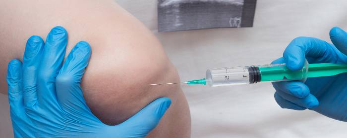
Orthopedic surgeons, rheumatologists, and other physicians have attempted to treat these conditions by injecting the damaged joint with one of the two main components of joint cartilage: extracellular matrix. Physicians inject hyaluronic acid (and sometimes related extracellular matrix proteins) to help replace and restore damaged joints. This approach can be helpful for some patients, but it is certainly not a cure.
Only recently, have researchers attempted to replace the other component of joint cartilage: chondrocytes. Specifically, researchers have focused their efforts on mesenchymal stem cells that have the ability to differentiate and become cartilage cells. Li and colleagues injected combinations of bone marrow-derived mesenchymal stem cells and hyaluronic acid into animals with experimental cartilage defects. They showed that hyaluronic acid injections alone modestly repaired the cartilage damage. However, when stem cells plus hyaluronic acid was injected, the joints were almost completely repaired. In other words, stem cells plus hyaluronic acid resulted in much greater improvement in joint cartilage damage than hyaluronic acid alone.
The authors of the study concluded that “bone marrow stem cells plus hyaluronic acid could be a better way to repair cartilage defects.” While additional work is needed, these results are extremely exciting for people who suffer from joint cartilage defects such as osteoarthritis. In the future, people who are candidates for hyaluronic acid injection treatments may instead receive a combination of hyaluronic acid plus stem cells and may enjoy an even greater benefit than hyaluronic acid treatment alone.
Reference: Li et al. (2018). Mesenchymal Stem Cells in Combination with Hyaluronic Acid for Articular Cartilage Defects. Scientific Reports. 2018; 8: 9900.

by admin | Jan 14, 2019 | Stem Cell Research, Stem Cell Therapy, Stroke
Patients who suffer ischemic stroke have some treatment
options, but many of them require immediate intervention and so are not useful
if too much time has elapsed between the stroke and treatment. Therapies that
employ stem cells are promising alternatives because stem cells can differentiate
into brain cells and potentially help to replace tissue that has been damaged
or destroyed.
A recent study published in Stem Cells
and Development has shown for the first time that a specific type of stem
cell – called ischemia-induced multipotent stem cells – may be able to help
with such repair of brain tissue in patients who have suffered a stroke.
Specifically, the research team demonstrated the technical ability to isolate
the ischemia-induced multipotent stem cells from the brains of elderly stroke
patients.
The scientists then used protein
binding techniques to determine where in the brain these stem cells came from.
They found that the cells came from areas of the brain where brain cells had been
damaged or killed from the stroke. These cells were located near blood vessels
and expressed certain biological markers that enabled the researchers to
confirm that they qualified as stem cells. Specifically, these cells had
proliferative qualities that suggested that they could potentially be used to
re-populate damaged areas of the brain. The cells also showed the ability to
differentiate into different types of cells, a key characteristic of stem cells
used for therapeutic purposes.
This study represents a
significant step in overcoming the technical challenges associated with
isolating and classifying ischemia-induced multipotent stem cells. The next
step for researchers will be to test the potential of these cells in stroke
treatment. If researchers show that these stem cells can be used to
successfully repair damaged areas of the brain – and more importantly, restore
functions that were disrupted by the stroke – then physicians and scientists
may be able to work together to translate these findings into therapies that
are regularly used in stroke.
Reference
Tatebayashi et al. 2017. Identification of multipotent stem
cells in human brain tissue following stroke. Stem Cells and Development, 26(11), 787-797.

by admin | Jan 4, 2019 | Stem Cell Research, Stem Cell Therapy, Studies
Spinal cord injury can be one of the most devastating
injuries. Long neurons that extend from the brain down the spinal cord are
severed and scarred. In most cases, this damage can never be repaired. If
patients survive an injury to the spinal cord, they can be permanently
paralyzed. Researchers have attempted to use high-dose steroids and surgery to
preserve the spinal cord, but these approaches are either controversial or
largely ineffective.
Ideally, one would create an environment in which nerve
cells in the spinal cord could regrow and take up their old tasks of sensation
and movement. One of the most promising approaches to do just this is stem cell
transplantation.
To test this concept, researchers used
stem cells derived from human placenta-derived mesenchymal
stem cell tissue (not embryonic stem cells) to form neural stem cells in
the laboratory. These neural stem cells have the ability to become neuron-like
cells, similar to those found in the spinal cord. The researchers then used
these stem cells to treat rats that had experimental spinal cord injury. The
results were impressive.
Rats treated with neural stem cells regained the partial
ability to use their hindlimbs within one week after treatment. By three weeks
after treatment, injured rats had regained substantial use of their hindlimbs.
The researchers confirmed that this improvement was due to neuron growth by
using various specialized tests (e.g. electrophysiology, histopathology). Rats
that did not receive stem cells did not regain substantial use of their
hindlimbs at any point in the study.
This work is particularly exciting because it shows that
stem cells can restore movement to animals who were paralyzed after spinal cord
injury. Moreover, the researchers used human stem cells derived from placenta,
which suggests that this effect could be useful in human spinal cord injury
patients (perhaps even more so than in rats). While additional work is needed,
these results offer hope to those who may one day develop severe spinal cord
injury.
Reference:
Zhi et al. (2014). Transplantation of placenta-derived
mesenchymal stem cell-induced neural stem cells to treat spinal cord injury.
Neural Regen Research, 9(24): 2197–2204.

by admin | Oct 30, 2018 | Studies, Stem Cell Research, Stem Cell Therapy
Spinal cord injury is the second leading cause of paralysis in the United States. When the spinal cord is severely injured, nerve cells in the spinal cord are damaged or destroyed. Also, a sort of scar forms in the affected area, which prevents nerve signals from traveling between the brain and the extremities. Consequently, people who sustain spinal cord injuries suffer from paralysis. The degree of paralysis depends on the location of the spinal cord injury; injuries higher on the spinal cord such as the neck or upper back area can lead to paralysis of all four limbs, for example. In almost all cases, the paralysis is permanent once it occurs, because nerve cells in the spinal cord do not regenerate.
Because spinal cord injuries are common and the consequences are usually permanent, researchers have been aggressively and tirelessly researching ways to treat this condition. One approach is to try to form new nerve cells in the spinal cord using stem cells. Mesenchymal stem cells can become new nerve cells given the right set of circumstances. Unfortunately, simply injecting mesenchymal stem cells into patients with severe spinal cord injuries cannot reverse paralysis. On the other hand, using exosomes from mesenchymal stem cells may be the push that stem cells need to become nerve cells in the spinal cord.
Exosomes are tiny packets of cellular material released by stem cells. They contain a variety of potentially beneficial substances; perhaps the most important in cell regeneration is micro RNA (miRNA). miRNA can cause complex changes in cells that simple drugs, proteins, or even regular RNA cannot. Researchers cannot easily deliver miRNA to where it is needed in the body, but exosomes taken from stem cells can deliver miRNA right where it needs to be.
Researchers collected human mesenchymal stem cells and placed them in an environment that would cause them to become nerve cells. But instead of simply using the stem cells directly, they instead collected the exosomes from those stem cells. Those exosomes could then be used to prompt mesenchymal stem cells to become nerve cells. Simply put, the exosomes drove the process more efficiently than the stem cells alone.
What does this all mean? Exosomes taken from the mesenchymal stem cells could eventually be used to treat spinal cord injury. Those special exosomes would magnify the nerve cell-creating effect, perhaps restoring nerve cell function to a damaged spinal cord. Considerable research needs to be done before this possibility becomes a clinical reality, but this knowledge helps researchers design targeted experiments in the future.

by admin | Oct 18, 2018 | Stem Cell Research, Stem Cell Therapy, Studies
A couple of weeks ago, scientists published findings showing that implanting human stem cells that are embedded within the engineered tissue can lead to the recovery of sensory perception in rats. The recovery of sensory perception is also accompanied by healing within the spinal cord and the ability to walk independently. The stem cells used in this experiment were collected from the membrane lining the mouth.
These results help demonstrate the potential for stem cells to help with spinal cord injuries but also point to the utility of combining stem cells with other factors to enhance their therapeutic effects. In this case, the researchers used a 3-dimensional scaffold to enable stem cells to attach and to stabilize them in the spinal cord. By adding growth factors, such as human thrombin and fibrinogen to the engineered tissue scaffolding, the researchers also increased the chances that attached stem cells would grow and differentiate.
The researchers compared the effects of their stem cell implants in paraplegic rats with the effects of adding no stem cells. Whereas the control rats who did not receive stem cells did not experience any improvement in mobility or sensation, 42% of the rats that did receive stem cells became better at supporting their weight on their hind limbs and at walking.
While these results are pre-clinical and do not apply directly to humans, the researchers conclude that further research is warranted. Given the positive impact of stem cells on the spinal cord in animals, it is reasonable to assume that stem cells may also benefit the human spinal cord. Further research will help clarify whether these stem cells can be adequately used to help treat patients with paraplegia.


 St. Petersburg, Florida
