Mesenchymal stromal cell therapy, often called MSC therapy, has become one of the most widely studied approaches in regenerative and cellular medicine. Over the past two decades, researchers have explored its potential to treat a wide range of inflammatory and immune-related conditions, including heart disease, lung injury, autoimmune disorders, and complications following cancer treatment.
As interest grows and more patients are invited to participate in clinical trials, one question becomes increasingly important: Is MSC therapy safe? This large, updated scientific review provides reassuring answers, demonstrating that MSC therapy continues to show a strong, favorable safety profile across thousands of patients and dozens of high-quality clinical trials.
As part of this review, Thompson et al. explain what MSC therapy is, why safety is such a critical concern, how researchers evaluated safety across many studies, and what the findings mean for patients, families, clinicians, and regulators.
Mesenchymal Stromal Cells and Their Therapeutic Role
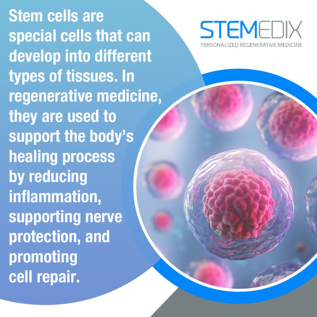
Mesenchymal stromal cells are multipotent cells that can be collected from adult tissues such as bone marrow, adipose tissue, and other sources. They were first described in the 1970s and have since gained attention for their ability to interact with the immune system and respond to inflammation.
Unlike embryonic stem cells, MSCs have a limited ability to turn into different cell types. Instead, their primary therapeutic value appears to lie in their ability to communicate with surrounding tissues. MSCs release bioactive molecules that help regulate immune responses, reduce excessive inflammation, and promote healing. They also migrate toward areas of injury or inflammation, making them attractive candidates for conditions where inflammation plays a central role.
Preclinical research, including animal studies, has shown promising results in conditions such as acute lung injury, sepsis, and heart attack. These findings have led to a growing number of human clinical trials evaluating both safety and potential benefits.
Safety Considerations for Mesenchymal Stromal Cell Therapy
Any therapy that involves living cells raises important safety questions. MSCs can divide, interact with the immune system, and circulate through the bloodstream. Because of these properties, scientists carefully monitor potential risks that could limit clinical use.
Early clinical trials suggested MSCs were generally safe, but these studies were small. As more trials were completed and patient numbers increased, researchers recognized the need to systematically review the evidence to identify any consistent safety signals that might not be obvious in individual studies.
Methods Used to Evaluate MSC Safety
To answer this question, researchers conducted a comprehensive systematic review and meta-analysis of randomized controlled trials. These trials compared patients who received MSC therapy via the bloodstream with those who received standard care or a placebo.
The review included studies published between 2012 and 2019 and built on an earlier 2012 review. The authors searched major medical databases and screened nearly 7,500 scientific papers. After careful evaluation, 55 randomized controlled trials involving 2,696 adult patients met the inclusion criteria.
By pooling data from multiple trials and using rigorous statistical methods, the researchers were able to estimate whether MSC therapy increased the risk of any negative events compared to control treatments.
Incidence of Fever Following MSC Therapy
Across all included trials, fever was the only adverse event that occurred more frequently in patients receiving MSC therapy compared to controls. Patients treated with MSCs were about 2.5 times more likely to develop a fever.
Notably, most of these fevers were mild and temporary. Although some fevers were reported as serious adverse events, they were rare overall. Fever typically occurred shortly after infusion and resolved without long-term consequences.
This finding is consistent with earlier reviews and is thought to reflect the body’s immune response to the infused cells rather than a sign of lasting harm. Recognizing fever as a known and manageable side effect helps clinicians monitor patients appropriately during and after treatment.
No Increased Risk of Infection, Thrombosis, or Malignancy
Beyond fever, the review found no significant increase in other major safety concerns. Patients receiving MSC therapy did not experience higher infection rates than controls, despite the cells’ immune-modulating effects. This suggests that MSCs do not meaningfully weaken immune defenses in clinical settings.
The analysis also found no association between MSC therapy and thrombotic or embolic events. Blood clots were rare overall and occurred at similar rates in both MSC-treated patients and control groups. This is particularly reassuring given ongoing research into how MSCs may interact with clotting pathways.
Perhaps most importantly, the review found no increased risk of malignancy. MSC-treated patients did not develop cancer more frequently than those in control groups. This finding addresses a longstanding concern related to the cells’ ability to proliferate and supports their continued investigation in clinical medicine.
Observed Reduction in Mortality
One of the more notable findings from this review was a reduced risk of death in patients receiving MSC therapy compared to controls. While this review was focused on safety rather than effectiveness, this observation suggests that MSC therapy does not increase mortality risk and may even offer protective benefits in specific patient populations.
It is important to interpret this finding cautiously, as the trials involved varied conditions and were not designed to measure survival as a primary outcome. Still, the absence of increased mortality provides further reassurance regarding safety.
Improved Safety Monitoring in Recent Trials
Compared to earlier studies, recent MSC clinical trials demonstrated improved attention to safety monitoring. More than three-quarters of the included trials reported having a predefined plan to track adverse events, a substantial improvement over earlier research.
Notably, none of the trials were stopped early due to safety concerns. Serious adverse events that were judged to be related or possibly related to treatment were extremely rare, occurring in only a small fraction of patients across all studies.
This progress reflects a maturing field that recognizes the importance of transparency, standardized reporting, and rigorous trial design.
Challenges in Trial Design and Reporting
Despite these encouraging findings, the authors highlighted areas that still need improvement. Only a small number of trials met all criteria for low risk of bias, indicating that study design quality varies widely across the field.
Reporting of MSC characteristics was another area of concern. Only a minority of trials fully described how MSCs were defined, tested for viability, or assessed for biological potency. These details are critical for understanding why some trials succeed while others do not.
Without consistent reporting standards, it becomes harder to compare results across studies or identify factors that influence outcomes. Improving transparency in cell characterization will be essential as newer, second-generation MSC products move into clinical trials.
Implications for Patients and Families
For patients considering participation in MSC clinical trials or learning about regenerative medicine options, this large body of evidence offers important reassurance. Across thousands of patients and dozens of trials, MSC therapy has consistently shown a strong safety record.
The most common side effect, fever, is generally temporary and manageable. Serious concerns such as infection, blood clots, cancer, and death have not been linked to MSC therapy when compared to standard treatments.
As with any investigational therapy, participation in clinical trials should involve careful discussion with healthcare providers, but concerns about safety alone should not be a barrier, given the current evidence.
Future Directions in MSC Research
MSC research continues to evolve as researchers learn more about how these cells work, how they interact with the immune and clotting systems, and how manufacturing methods influence their behavior. Future trials will explore new indications, refined dosing strategies, and enhanced cell products designed to improve consistency and effectiveness.
Ongoing safety monitoring remains essential, particularly as therapies move into larger and more diverse patient populations. Continued adherence to rigorous trial design and transparent reporting will help ensure that advances in regenerative medicine are both effective and safe.
Overall Conclusions on MSC Safety
Thompson et al.’s updated review provides the most comprehensive evaluation to date of the safety of mesenchymal stromal cell therapy in adult clinical trials. Aside from an increased likelihood of fever, no meaningful safety signals were identified across thousands of patients.
The findings reinforce the conclusion that MSC therapy continues to demonstrate a favorable safety profile. For researchers, clinicians, regulators, and patients alike, this growing body of evidence supports the responsible, ongoing development of MSC-based therapies as part of the evolving field of regenerative medicine.
As research progresses, maintaining high standards for study design, cell characterization, and adverse event reporting will be key to translating this promising therapy into broader clinical practice.
Source: Thompson M, Mei SHJ, Wolfe D, Champagne J, Fergusson D, Stewart DJ, Sullivan KJ, Doxtator E, Lalu M, English SW, Granton J, Hutton B, Marshall J, Maybee A, Walley KR, Santos CD, Winston B, McIntyre L. Cell therapy with intravascular administration of mesenchymal stromal cells continues to appear safe: An updated systematic review and meta-analysis. EClinicalMedicine. 2020 Jan 17;19:100249. doi: 10.1016/j.eclinm.2019.100249. PMID: 31989101; PMCID: PMC6970160.
Mesenchymal stromal stem cells, commonly called MSCs, have been among the most-studied cell types in regenerative medicine over the past two decades. They have been tested in hundreds of clinical trials for conditions ranging from joint degeneration to heart disease, autoimmune disorders, lung injury, and complications after transplantation.
MSCs have consistently been shown to be safe, but their effectiveness has been mixed. Many trials have not met their main efficacy goals, and only a small number of MSC-based products have received regulatory approval worldwide.
This review by Lu and Allickson examines what has been discovered about MSC therapy and what remains to be done before these therapies can be widely adopted in routine clinical practice.
From Bone Marrow Cells to Powerful Immune Modulators
MSCs were first identified in mouse bone marrow as cells that could support blood-forming stem cells and form bone, cartilage, and fat. Human MSCs were later isolated in the 1990s. Early on, much of the excitement around MSCs focused on their ability to turn into different mesodermal tissues and directly replace damaged cells.
However, over time, it became clear that this “replacement” model did not fully explain what was happening in living organisms. In patients, MSCs do not routinely transform into large amounts of new tissue. Instead, their main therapeutic effects appear to come from the signals they send out rather than the cells they become.
Today, most researchers view MSCs as “medicinal signaling cells.” They can self-renew and still form bone, cartilage, and muscle, but their real power lies in their paracrine effects. MSCs sense damage and inflammation in their environment and respond by releasing a complex mix of biologically active molecules. This includes cytokines, chemokines, growth factors, extracellular matrix components, and extracellular vesicles that carry proteins, lipids, and genetic material, such as microRNAs. These signals help guide other cells to repair tissue, grow new blood vessels, calm harmful immune responses, and limit scarring.
How MSCs Influence Repair and Immunity
MSCs have been shown in laboratory and animal studies to home to sites of injury and support tissue repair in the heart, lungs, joints, nervous system, and other organs. They create a local microenvironment that encourages healing, reduces cell death, and can improve organ function after injury.
Equally important is their role in immune modulation. MSC-derived factors can shift the immune system away from a highly inflammatory state and toward a more balanced, regulatory profile. They interact with many types of immune cells, including T cells, B cells, macrophages, dendritic cells, and natural killer cells, and can either dampen or support immune activity depending on the context. This flexible, environment-dependent behavior is one of the reasons MSCs are being studied for such a wide range of inflammatory and immune-mediated conditions.
Extracellular vesicles released by MSCs, also known as MSC-derived EVs, are a significant contributor to their effectiveness. These tiny membrane-bound packages carry proteins, RNAs, and other molecules that can travel to distant cells and influence their behavior. EVs from MSCs have shown the ability to reduce fibrosis, promote tissue regeneration, and calm inflammation in preclinical models, raising interest in EVs as a possible “cell-free” therapy that might someday complement or even replace live cell treatments.
Defining an MSC: Why Standards Matter
One ongoing challenge in MSC research is that not all MSCs are the same. They can be derived from many different tissues, including bone marrow, adipose tissue, and perinatal tissues such as the placenta and the umbilical cord. Each source can produce cells with different characteristics, and even cells from the same source can vary based on how they are collected, cultured, and stored.
To create consistency in the field, the International Society for Cellular Therapy established basic criteria in 2006 to define human MSCs. According to these guidelines, MSCs must adhere to plastic in standard lab cultures, express specific surface markers, and differentiate into bone, cartilage, and fat cells under appropriate laboratory conditions.
Even with these guidelines, the authors note that there remains considerable variability across MSC products. Differences in cell source, donor characteristics, manufacturing methods, dosing strategies, and delivery routes all contribute to the wide range of outcomes seen in clinical trials. This variability is one of the main reasons it has been difficult to draw simple conclusions about “MSC therapy” as a single, uniform treatment.
Regulatory Approvals: A Few Successes Among Many Trials
Despite the large number of registered MSC trials worldwide, only a limited number of MSC-based products have received regulatory approval so far. Different countries regulate cell therapies through agencies similar to the U.S. Food and Drug Administration, such as Health Canada, the European Medicines Agency, and others in Asia.
One important milestone highlighted in this review is the recent approval in the United States of an MSC therapy for pediatric graft-versus-host disease, a serious complication of stem cell transplantation. This marks the first MSC therapy approved by the FDA and demonstrates that, under the right conditions, MSCs can meet the rigorous safety, quality, and benefit standards required by regulators.
Outside the U.S., several other MSC-based products have been approved for conditions such as cartilage defects and graft-versus-host disease. However, when viewed against the backdrop of hundreds of trials, the number of approvals remains small, emphasizing how challenging it has been to translate the promise of MSCs into consistent, reproducible clinical benefit.
What the Clinical Trial Landscape Looks Like
A recent search of the ClinicalTrials.gov database found hundreds of registered studies involving mesenchymal stromal or mesenchymal stem cells, covering early-phase safety trials through more advanced phase 3 and 4 studies. These trials span a wide range of indications, from orthopedic and cardiovascular disorders to autoimmune diseases, neurological conditions, and complications of cancer treatment.
Yet, a key concern is that the vast majority of these trials have not reported their results publicly. This lack of accessible outcome data makes it difficult for clinicians and researchers to fully understand where MSCs are working well, where they are not, and what factors may explain the differences. It also slows progress in refining protocols and designing better future studies.
Safety: A Clear Strength of MSC Therapy
One consistent and reassuring theme across the MSC literature is safety. Clinical trials over more than two decades have shown that MSC therapy is generally very well tolerated. Reports of serious infusion reactions, organ damage, severe infections, cancers, or treatment-related deaths directly attributable to MSCs have been extremely rare.
Safety data is especially strong for bone marrow–derived and adipose-derived MSCs, which have the longest track record in human studies. Newer sources, including perinatal tissues, also appear promising but may benefit from longer follow-up and more comprehensive monitoring as experience grows.
The Efficacy Challenge and Future Directions
While safety has been firmly established, efficacy has been much less consistent. Many MSC trials have failed to meet their primary endpoints, and in some cases, the benefits have been modest or difficult to distinguish from placebo or standard care. This is not unique to MSCs—many new therapies face similar hurdles—but it does mean that expectations must be realistic.
Lu and Allickson emphasize that the next chapter for MSC therapy will depend on solving several key problems. These include better defining which patients and diseases are most likely to respond, standardizing and optimizing cell manufacturing, clarifying dose and timing, and understanding how factors like age, comorbidities, and prior treatments influence outcomes. It will also be important to determine when MSCs should be used alone and when they may be most effective in combination with other therapies.
What This Means for Patients Today
The data shows that MSCs are safe with clear potential for tissue repair and immune modulation. At the same time, the field is still working to consistently translate these biological effects into strong, repeatable clinical benefits across many diseases.
As research continues, mesenchymal stromal cell therapy remains one of the most carefully studied and promising avenues in regenerative medicine. The progress to date provides a strong foundation, and the future outlook will depend on rigorous science, thoughtful trial design, and continued collaboration between researchers, clinicians, regulators, and patients.
If you or someone you care about has been diagnosed with a spinal cord injury, you understand how life-altering the challenges can be. At Stemedix, we work with patients who have already received a confirmed diagnosis and are seeking alternative ways to support their recovery goals. While no treatment guarantees a cure, regenerative medicine offers the potential to support healing and reduce the impact of symptoms through biologically active therapies.
Stem cell therapy for spinal cord injury is one such approach that may help promote cellular repair, reduce inflammation, and encourage nerve support. You won’t find exaggerated claims or comparisons here, just realistic, patient-focused information backed by experience. We customize each treatment plan using the documentation you provide, and we support you throughout your journey. This article will walk you through the basics of spinal cord injury, explain how stem cells for the treatment of spinal cord injury are used, and outline what to expect with our process.
What is Spinal Cord Injury?
A spinal cord injury (SCI) is damage to the spinal cord that disrupts communication between the brain and the body. When this pathway is damaged, the body’s ability to send and receive signals becomes impaired. That can mean a loss of movement, sensation, or automatic functions like bladder and bowel control. Most spinal cord injuries happen because of sudden trauma. Studies show that the most common causes of SCI were automobile crashes (31.5%) and falls (25.3%), followed by gunshot wounds (10.4%), motorcycle crashes (6.8%), diving incidents (4.7%), and medical/surgical complications (4.3%).
The spinal cord does not regenerate the way some tissues in the body do. This makes the injury permanent in many cases. The outcome depends on where the injury occurred and how much of the nerve pathway is still intact.
Types and Locations of Spinal Cord Injuries
Spinal cord injury (SCI) is classified by severity, complete or incomplete, and by the spinal region affected. A complete injury results in loss of all movement and sensation below the injury site, while incomplete injuries allow some function. The spinal region involved guides recovery and therapy goals.
Cervical nerve injuries (C1–C8) impact the neck, arms, hands, and breathing, with higher levels possibly requiring ventilation support. Thoracic injuries (T1–T12) affect chest and abdominal muscles, impacting balance and trunk control. Lumbar and sacral injuries (L1–S5) influence leg movement and bladder function, with outcomes varying based on injury extent and completeness.
Common Symptoms and Challenges After SCI
Patients with SCI may experience paralysis, sensory loss, chronic pain, and complications in daily functions. Spinal cord injury affects more than movement. Many patients deal with muscle spasticity, pressure injuries due to immobility, frequent urinary tract infections, and problems with body temperature control. Autonomic dysreflexia, a sudden increase in blood pressure triggered by stimuli below the injury level, is a serious risk in those with injuries at or above T6. Emotional and psychological responses, including anxiety and depression, are also common and require support.
At Stemedix, we recognize that each spinal cord injury is unique. We tailor every treatment plan based on the medical records and information you provide, not generalized assumptions. If you’re exploring stem cells for the treatment of spinal cord injury, our team is ready to walk you through options that align with your health history and functional goals.
What is Regenerative Medicine?
Regenerative medicine supports the body’s repair mechanisms by introducing biologically active materials. This field focuses on helping your body respond to damage by using living cells and biological components. Instead of masking symptoms, regenerative treatments aim to influence the cellular environment that surrounds the injured tissue. In many cases, this includes the use of stem cells and growth factors.
For individuals with a spinal cord injury, regenerative medicine introduces new options that may encourage healing responses the body struggles to activate on its own. While this type of therapy doesn’t replace rehabilitation, it may work alongside your current efforts to promote tissue stability and reduce secondary complications.
Stem Cell Therapy as a Treatment Option for SCI
Stem cell therapy for spinal cord injury is being explored to support recovery and symptom relief. Researchers are investigating how stem cells may influence the biological environment of an injured spinal cord. You won’t find a generalized approach here. Stem cell treatment for spinal cord injury is tailored to each case based on the location of injury, severity, and medical history.
The focus is not on reversing the damage or offering a cure. Instead, stem cells for the treatment of spinal cord injury may help by releasing chemical signals that support the health of nearby nerve cells, protect against further breakdown, and potentially stimulate limited repair processes. Some patients have reported improvements in muscle control, sensation, or bladder regulation, though outcomes vary and remain under study.
How Stem Cells Work to Support Healing
Stem cells can develop into specialized cell types and secrete proteins that support tissue repair. These cells have two key roles in regenerative medicine. First, they can adapt to different cell types, such as those found in the nervous system. Second, and equally important, they release helpful proteins, like cytokines and growth factors, that create a healing-friendly environment. This may reduce chronic inflammation and improve communication between nerve cells that remain intact.
In spinal cord injury cases, these cells may influence glial scar formation, improve blood flow to the damaged region, and protect vulnerable cells from oxidative stress. For example, studies have shown that transplanted mesenchymal stem cells can release brain-derived neurotrophic factor (BDNF), which plays a role in supporting neural survival.
At Stemedix, we offer regenerative therapy based on the existing diagnosis and medical documentation provided by each patient. Our approach respects the experimental nature of this therapy while offering guidance and structure throughout the process.
Potential Benefits of Stem Cell Therapy for Spinal Cord Injury
Exploring the potential benefits of stem cell therapy gives you a chance to learn how regenerative medicine may support certain aspects of your spinal cord injury recovery. While results vary for each individual, many patients report improvements in pain, movement, and physical function over time.
Pain Reduction and Muscle Relaxation
Many patients report decreased neuropathic pain and reduced muscle tension following therapy. Neuropathic pain is one of the most common and challenging symptoms following spinal cord injury. You may experience burning, tingling, or shooting sensations due to misfiring nerves. For some individuals receiving stem cell therapy for spinal cord injury, these symptoms become less intense or more manageable. This could be related to how certain types of stem cells interact with immune cells and inflammatory pathways.
Studies have suggested that mesenchymal stem cells (MSCs), for example, can release bioactive molecules that influence the environment surrounding injured nerves and even interact with neural cells in spine and brain conditions. In some cases, patients also describe less spasticity or tightness in the muscles, which can reduce discomfort during sleep or daily movement.
Improved Circulation and Motor Function
Stem cell treatment for spinal cord injury may support vascular health and contribute to smoother movement. Reduced blood flow after a spinal cord injury can limit your body’s ability to heal or respond to therapy. You might notice cold extremities, swelling, or slower wound healing. Stem cell therapy may support microvascular repair by promoting angiogenesis, the formation of new blood vessels in damaged tissues. This improved circulation helps deliver oxygen and nutrients more efficiently to the affected areas. Some individuals receiving stem cell therapy report smoother joint movement, greater control over posture, and better balance during transfer or mobility tasks.
Increased Muscle Strength and Abilities
Muscle engagement and strength may increase as nerve signals improve. After a spinal cord injury, the connection between your brain and muscles may be disrupted or weakened. Over time, this can lead to muscle wasting or limited control. For individuals receiving stem cell treatment for spinal cord injury, some report noticeable changes in muscle tone, voluntary movement, or strength, especially in the lower limbs or core. These observations tend to occur in cases where some nerve pathways remain intact.
For example, a patient with an incomplete thoracic injury might regain the ability to perform assisted standing exercises or show improvements in hip stability. While not every case leads to increased muscle output, any gains in strength can contribute to mobility training, sitting tolerance, and daily activities.
Patient Experience and Reported Outcomes
Individuals receiving therapy frequently describe improvements in mobility, energy levels, and daily activity. Each patient arrives with unique goals. Some hope to walk again. Others want to reduce fatigue or rely less on medications. After therapy, individuals often share changes that impact their quality of life, such as being able to transfer with less assistance, participate in treatment longer, or sleep more comfortably.
At Stemedix, we focus on your specific history, symptoms, and expectations before building a treatment plan. These outcomes help us communicate realistic possibilities, while always making it clear that regenerative medicine is still considered experimental.
How Stemedix Approaches Stem Cell Therapy for SCI
Every individual with a spinal cord injury has a different medical background and a different journey. That’s why your treatment experience with Stemedix begins with your history, not just your condition.
Customized Treatment Based on Patient History
Stemedix develops treatment plans based on medical records submitted by the patient. If you’ve already received a spinal cord injury diagnosis, our team starts by reviewing the medical documents you send us. This includes imaging studies, physician assessments, and any other relevant details about your injury. By focusing on those who have already completed a diagnostic evaluation, we’re able to provide a more appropriate regenerative therapy experience.
We do not perform physical exams or order MRIs. If your current records are outdated, we can help gather updated information on your behalf once you sign a simple medical release form. This makes sure that our team has the most accurate data to tailor a regenerative approach based on your unique condition, designing therapy around what your body truly needs, not generalized assumptions.
Role of Board-Certified Physicians and Care Coordinators
Each case is reviewed by board-certified physicians experienced in regenerative medicine. When you choose to move forward, your medical information is assessed by physicians who specialize in regenerative therapies. They have experience working with spinal cord injury patients and understand how stem cell therapy may support certain biological functions involved in healing.
Patients are supported by dedicated Care Coordinators who handle logistics, scheduling, and communication. You won’t be left navigating the details alone. Once your evaluation is underway, a Care Coordinator will work closely with you to keep the process on track. This includes walking you through the next steps, answering questions, and helping schedule your treatment. Having one point of contact makes the entire journey easier to follow and less overwhelming.
Patient Support Services and Accommodations
Stemedix offers assistance with travel arrangements, transportation, and medical support equipment. Whether you’re located nearby or traveling across the country, we help remove logistical barriers. Our team can coordinate hotel stays, provide complimentary ground transportation, and arrange for wheelchair-accessible options if needed.
Whether a patient is local or traveling from another state, Stemedix helps coordinate hotels and driver services to make the process more accessible. Your focus should be on preparing for therapy, not stressing over logistics.
Getting Started with Stemedix
How to Connect with a Care Coordinator
Our Care Coordinators are ready to assist you at every step. They can answer your questions, review your medical documents, and guide you through the application process. From your initial inquiry through follow-up care, they provide consistent support to help you understand the next steps in pursuing stem cell therapy for spinal cord injury.
What to Expect During the Treatment Process
Once your case is reviewed and approved by our physicians, you will receive a customized treatment plan with a scheduled date for your therapy. Treatment is provided in a licensed medical facility under the supervision of experienced professionals. After treatment, ongoing follow-up is available to monitor your progress and provide additional support as needed.
Contact Stemedix Today
If you are interested in learning more about stem cell treatment for spinal cord injury, request an information packet today. The team at Stemedix is here to guide you on your journey to better health. Call us at (727) 456-8968 or email yourjourney@stemedix.com to know more.
Autoimmune and rheumatic diseases affect millions of people worldwide and can involve the joints, skin, gut, nervous system, and many other organs. Conditions like rheumatoid arthritis, osteoarthritis, lupus, inflammatory bowel disease, multiple sclerosis, and Sjögren’s syndrome often cause chronic pain, fatigue, and progressive damage.
Standard treatments usually focus on calming the immune system with medications such as steroids, immunosuppressants, or biologic drugs. While these can be effective, they often come with side effects, do not work for everyone, and rarely offer a true long-term cure.
Because of this, there is growing interest in therapies that can not only reduce inflammation but also help reset the immune system and support tissue repair.
Mesenchymal stem cells (MSCs) are among the most studied cell types in this field. An extensive new analysis of randomized controlled trials focused specifically on MSCs for autoimmune and rheumatic diseases to better answer two key questions: how well do they work, and how safe are they?
Understanding Mesenchymal Stem Cells and Their Role in Immune Disease
Mesenchymal stem cells are a type of adult stem cell that can be isolated from many tissues, including bone marrow, adipose tissue, umbilical cord, placenta, and dental pulp. They can renew themselves, form bone, cartilage, and fat cells under certain conditions, and, significantly, interact with the immune system.
MSCs have low expression of surface markers that typically trigger rejection, so they can often be used from a donor without provoking a strong immune response. They can also “sense” inflammatory signals and respond by releasing a range of anti-inflammatory and tissue-supporting molecules. These include cytokines, growth factors, and extracellular vesicles that can influence T cells, B cells, macrophages, dendritic cells, and other immune system influencers.
Because of these properties, MSCs are being studied as a way to calm overactive immune responses, promote immune tolerance, and support repair in tissues damaged by chronic inflammation. Researchers are exploring their potential as an add-on or alternative to traditional immunosuppressive therapies in many autoimmune and rheumatic conditions.
How the Study Was Conducted
To get a clearer picture of MSCs across diseases, Zeng et al. performed a systematic review and meta-analysis of randomized controlled trials. They searched major English and Chinese medical databases through December 2023 and identified 42 randomized controlled trials involving 2,183 participants.
These trials covered several autoimmune and rheumatic conditions, including rheumatoid arthritis, osteoarthritis, spondyloarthritis, systemic sclerosis, systemic lupus erythematosus, inflammatory bowel disease, multiple sclerosis, and primary Sjögren’s syndrome.
The research team looked at how MSC therapy affected symptoms, disease activity scores, pain scales, and other clinical measures in each disease. They also carefully examined safety by comparing rates of adverse events, such as infections, worsening disease, and other complications, between MSC-treated patients and control groups.
Overall Findings: Promising Benefits in Some Diseases, Mixed Results in Others
The conclusion from this analysis is that MSC therapy shows encouraging benefits in several autoimmune and rheumatic diseases, while in others, the results are more modest or still unclear.
For osteoarthritis, MSC injections into the joint were associated with meaningful improvements in pain and function. Across multiple trials, patients who received MSCs from bone marrow, umbilical cord, or adipose tissue reported less pain on visual analog scales and better scores on standard osteoarthritis questionnaires, particularly in the adipose-derived MSC group. Stiffness did not consistently improve, but overall pain and function did.
In systemic lupus erythematosus, MSC therapy led to significant reductions in disease activity scores and improvements in kidney-related measures such as protein in the urine. Complement levels, which are often low in active lupus, improved in the MSC-treated group. These changes suggest a significant impact on the underlying immune activity, not just symptoms.
In inflammatory bowel disease, including Crohn’s disease and ulcerative colitis, MSC therapy improved clinical response and remission rates compared to control treatments. This aligns with previous work showing benefits in challenging cases, such as complex perianal fistulas.
By contrast, in multiple sclerosis and systemic sclerosis, the meta-analysis did not show a clear improvement in key outcomes such as lesion number or disability scores for MS, or consistent, statistically strong benefits for SSc. That does not mean there is no benefit at all; it may reflect limited trial numbers, small sample sizes, or the need for more optimized treatment protocols.
For conditions like rheumatoid arthritis, spondyloarthritis, and primary Sjögren’s syndrome, the results are encouraging but still based on relatively few randomized trials. Early studies suggest improvements in pain, function, disease activity scores, and gland function, but larger, longer-term trials are needed.
Key Findings Across Individual Diseases
In patients with knee osteoarthritis, intra-articular MSC injections improved pain and physical function. Patients who received MSCs, especially from adipose tissue, reported better walking ability, reduced discomfort, and overall improved joint function. Although cartilage regeneration is still being actively studied, these results support MSCs as a potential tool for symptom relief and functional improvement.
In rheumatoid arthritis, a small number of trials showed that bone marrow–derived MSCs were well tolerated and associated with reduced disease activity, better joint symptoms, and meaningful response rates that lasted up to a year in many patients. Immunologic measures also shifted in a more favorable direction, with reduced inflammatory signals.
In spondyloarthritis, early data from a single randomized trial suggest possible improvement compared to a standard biologic treatment, but the evidence base is still very limited.
In systemic sclerosis, one trial using adipose-derived regenerative cells suggested some improvement in hand function and disability scores in patients with diffuse cutaneous disease, especially over longer follow-up, but not all results reached statistical significance.
In primary Sjögren’s syndrome, MSC therapy improved dryness symptoms, salivary and tear gland function, and reduced disease activity scores in the trial included in this review. Laboratory markers such as immunoglobulin levels and inflammatory markers also improved.
In systemic lupus erythematosus, MSCs reduced disease activity and improved kidney involvement, while maintaining a safety profile similar to standard therapy.
In inflammatory bowel disease, MSC therapy improved clinical efficacy without raising the rate of adverse events, supporting its role as a potential option, particularly in complex or treatment-resistant cases.
In multiple sclerosis, MSC therapy did not significantly improve lesion counts, lesion volume, or disability scores in the combined analysis of randomized trials. However, many early-phase, non-randomized studies still support the safety of MSCs and suggest potential benefits that need confirmation in better-designed, larger trials.
Safety Findings: No Increase in Adverse Events
One of the most important questions for any new therapy is safety. In this extensive review, MSC transplantation did not increase the risk of adverse events in the conditions studied.
For osteoarthritis, lupus, inflammatory bowel disease, and multiple sclerosis, the rates of adverse events were similar between MSC-treated patients and control groups. In other words, adding MSC therapy did not make side effects more common. Notably, there was no signal that MSCs increased serious risks such as infections, malignancy, or severe treatment-related complications across these trials.
This supports the idea that MSC therapy, when prepared and administered under appropriate clinical protocols, has a favorable safety profile in autoimmune and rheumatic diseases. However, as with any treatment, patients should be monitored carefully, and long-term follow-up remains essential.
Immune Regulation by MSCs in Autoimmune and Rheumatic Disease
Although each disease is different, many autoimmune and rheumatic disorders share a common theme: the immune system loses tolerance and begins attacking the body’s own tissues. MSCs seem to help by gently “rebalancing” the immune system rather than shutting it down completely.
MSCs can reduce overactive T and B cell responses, promote regulatory T cells that help maintain tolerance, and shift inflammatory cells toward more calming, tissue-protective roles. They also release factors that support tissue repair, improve the local microenvironment, and influence pathways involved in healing and regeneration.
This multi-layered action may explain why MSCs show promise across different diseases that all have an immune and inflammatory component, even though the specific organs affected are not the same.
Remaining Challenges and Future Directions
Despite promising signals, the authors of the review emphasize that MSC therapy is not a one-size-fits-all solution and that there is still work to be done. Different studies used different cell sources, doses, timing, and treatment schedules. These differences likely contribute to the variation in results.
The researchers also suggest that MSCs are most likely to be effective when combined with other treatments rather than used alone, that early intervention may be more beneficial than late-stage treatment, and that multiple doses may be more effective than a single infusion in some cases. They also stress the importance of tailoring protocols to the specific disease and patient rather than applying a rigid standard formula.
Larger, high-quality randomized controlled trials are still needed, especially in conditions like rheumatoid arthritis, spondyloarthritis, systemic sclerosis, multiple sclerosis, and primary Sjögren’s syndrome, where early results are promising but not yet definitive.
What These Findings Mean for Patients
For patients and families living with autoimmune or rheumatic immune diseases, this analysis offers cautious optimism. The evidence suggests that mesenchymal stem cell transplantation may help reduce symptoms and disease activity in several conditions, especially osteoarthritis, systemic lupus erythematosus, inflammatory bowel disease, and primary Sjögren’s syndrome, with encouraging signals in rheumatoid arthritis and some others.
Just as importantly, MSC therapy appears to have a favorable safety profile in the clinical trials analyzed, with no increase in overall adverse events compared to standard treatments or placebo.
However, MSC therapy is still being actively studied, and it is not yet a universally established standard of care for these diseases.
As research continues, the goal is to refine MSC-based treatments so they are safer, more consistent, and more effective, helping address the unmet needs of people living with chronic autoimmune and rheumatic diseases.
Source: Zeng, L., Liu, C., Wu, Y. et al. Efficacy and safety of mesenchymal stromal cell transplantation in the treatment of autoimmune and rheumatic immune diseases: a systematic review and meta-analysis of randomized controlled trials. Stem Cell Res Ther 16, 65 (2025). https://doi.org/10.1186/s13287-025-04184-x
Low back pain is one of the most common health problems worldwide. It affects quality of life, limits work and daily activities, and creates a significant economic burden. In many adults, especially those over age 50, a key driver of this pain is lumbar intervertebral disc degeneration. When the disc between the vertebrae begins to break down, it can become a source of chronic, deep “discogenic” pain that is often difficult to treat.
Traditional treatment options include physical therapy, medications, injections, and, in some cases, surgery. These treatments can help manage symptoms but do not always address the underlying disc damage, and surgery is not suitable or desirable for everyone. This is why researchers have been exploring regenerative approaches, including mesenchymal stem cell (MSC) therapy, to repair or stabilize the disc and reduce pain at its source.
A recent review and meta-analysis by Zhang et al. examined the effectiveness of MSC injections into the disc for patients with lumbar discogenic pain and whether this approach is safe. The results are promising and add to the growing body of evidence supporting MSC-based therapies for spine-related conditions.
Why Disc Degeneration Causes Low Back Pain
The intervertebral discs act as shock absorbers between the vertebrae in the spine. Each disc has a soft, gel-like center (the nucleus pulposus) surrounded by a tougher outer ring (the annulus fibrosus). Over time, age, genetics, mechanical stress, and lifestyle factors can lead to degeneration of these discs. The disc can lose water content, become thinner, and develop small tears.
When this happens in the lumbar spine, it can trigger discogenic low back pain. This type of pain often feels deep, aching, and persistent. It may worsen with sitting or bending and improve when lying down.
Initial treatment typically involves non-surgical approaches such as exercise therapy, manual therapy, nonsteroidal anti-inflammatory drugs, and other pain-modulating medications. While many patients improve, others continue to have significant pain and disability even after trying conservative treatments for months or years.
How Mesenchymal Stem Cells May Help Degenerative Discs
Mesenchymal stem cells are a type of adult stem cell that can be obtained from bone marrow, adipose tissue, cartilage, and other sources. They are known for several beneficial properties. Under the right conditions, MSCs differentiate into bone, cartilage, and other mesenchymal tissues under the right conditions. They secrete a variety of growth factors and signaling molecules that support tissue repair and modulate inflammation. They also communicate directly with nearby cells to influence the local environment.
In the context of disc degeneration, the idea is to inject MSCs directly into the damaged disc. Once there, they may help repopulate the disc with healthier cells, support the remaining disc cells, and alter the inflammatory and degenerative microenvironment.
According to the authors, animal studies and early human trials have suggested that MSC injections into degenerated discs can improve disc hydration, reduce pain, and enhance function. However, these individual clinical studies tend to be small and vary in design, making it hard to draw firm conclusions from any single trial. This is where a meta-analysis, which pools data from multiple studies, becomes particularly valuable.
How the Meta-Analysis Was Conducted
For this meta-analysis, researchers reviewed several major medical databases, including PubMed, Web of Science, Embase, and the Cochrane Library, through September 18, 2022. They focused on clinical studies that examined MSC treatment for lumbar disc degeneration and disc-related low back pain.
Of the 2,392 studies initially identified, 9 met the inclusion criteria. These studies included 245 patients, most of whom received injections of bone marrow–derived MSCs directly into damaged discs. Study quality was evaluated using the Newcastle–Ottawa Scale, and standard meta-analysis methods were used to analyze the data.
The primary outcomes measured were changes in pain levels and changes in the Oswestry Disability Index (ODI), which assesses how back pain affects daily activities. Researchers also reviewed reoperation rates and side effects to evaluate safety.
Pain Relief: Improvements on the Visual Analogue Scale
Pain was measured using the Visual Analogue Scale (VAS), where patients rated their pain on a simple numerical scale. Across the studies, patients who received MSC injections showed clear reductions in pain from the start of treatment to the final follow-up.
When the data were combined, average pain scores improved by more than 40 points on a 0–100 scale. This represents a significant and meaningful decrease in pain for many patients. Although results varied between studies, nearly all showed pain improvement with MSC treatment.
Other meta-analyses have reported similar findings, showing that MSC therapy can significantly reduce pain in people with disc degeneration. This analysis supports those results and suggests that MSC injections provide meaningful pain relief for appropriately selected patients.
Improved Function and Reduced Disability: Oswestry Disability Index Results
Pain is only part of the issue. For many people with chronic lumbar discogenic pain, the most important question is whether they can get back to their everyday lives. This is where the Oswestry Disability Index (ODI) was especially helpful.
In the meta-analysis, ODI scores improved significantly after MSC injection. The pooled data showed an average improvement of more than 20 points from baseline to the final follow-up, indicating better function and less disability. This means that patients were not only reporting less pain, but they were also better able to sit, stand, walk, work, and perform self-care.
Taken together, the pain and disability findings suggest that MSC therapy has the potential to provide both symptom relief and functional benefit in patients with disc-related low back pain.
Safety and Reoperation Rates: A Reassuring Profile
Any new therapy needs to be evaluated not just for benefit but also for risk. In this meta-analysis, MSC injection therapy for discogenic low back pain demonstrated a favorable safety profile.
No serious adverse events related to the MSC therapy were reported across the included studies. Treatment-emergent side effects, when they occurred, were generally mild and included symptoms such as back, joint, or muscle pain, which are also common in the underlying condition. Previous meta-analyses in this area have similarly reported no statistically or clinically significant increase in adverse events with MSC injections.
The pooled reoperation rate was low, around 7%. This suggests that most patients did not require further surgical intervention at the treated level during follow-up. While longer-term data are still needed, the findings support the idea that MSC disc injections are both safe and potentially protective against the need for additional procedures in the short- to medium-term.
MSCs Compared With Other Cell-Based Strategies for Disc Repair
MSCs are not the only cell type being studied for disc repair. Disc-derived chondrocytes and nucleus pulposus cells have also been explored. These cells can be harvested from disc tissue, expanded in the lab, and reimplanted. However, this approach has challenges. Disc cells have a limited natural capacity to multiply, and obtaining enough cells may require harvesting from other discs, which can be invasive and may compromise healthy tissue.
According to the authors, MSC-based therapies offer several advantages. MSCs can be isolated from bone marrow, adipose tissue, and other sources and expanded in culture to achieve therapeutic doses. Bone marrow–derived MSCs, used in all nine influential clinical studies in this meta-analysis, can differentiate into nucleus pulposus-like cells and support existing disc cells by secreting beneficial cytokines, such as transforming growth factor-beta 1.
That said, bone marrow harvest is invasive and yields relatively few MSCs. Adipose-derived MSCs, which can be obtained in higher quantities from fat tissue, are an attractive alternative and may have strong anti-inflammatory properties. Adipose tissue, which naturally contains MSCs, has shown promising results in joint applications and is being explored for discogenic pain, although these data were not included in the current analysis. This highlights that most of the evidence so far is for bone marrow–derived MSCs, and more research is needed on other sources.
Limitations of the Current Evidence
As encouraging as the results are, it is essential to interpret them in context. Zhang et al. point out that the meta-analysis has several limitations. The number of clinical studies and the total number of patients remain relatively small. Of the 245 patients included, 193 received bone marrow–derived MSC injections, limiting the ability to generalize the findings to all MSC products.
Not all studies reported pain and disability outcomes in the same way. Only four studies provided complete VAS data suitable for pooled analysis, and only five contributed to the ODI analysis. Differences in how scales were reported and in follow-up timing can introduce variability and make it harder to fully capture the treatment effect.
Additionally, most of the included studies focused on single-level disease and carefully selected patients. Outcomes in broader, more varied patient populations may differ. Longer-term data are also needed to determine how durable the benefits are and whether MSC therapy can truly halt or reverse disc degeneration over many years.
Finally, this analysis focuses on bone marrow–derived MSCs and does not fully address other MSC sources such as adipose tissue, synovium, or perinatal tissues. Future trials will be needed to compare cell sources, doses, and delivery methods more systematically.
What This Means for Patients With Discogenic Low Back Pain
For patients living with chronic, disc-related low back pain that has not improved with standard conservative care, this meta-analysis offers cautious optimism. The pooled data suggest that MSC injections into the degenerated disc can significantly reduce pain and improve function, with a low rate of serious side effects and reoperations.
MSC therapy is not yet a universal, first-line treatment for discogenic pain, and much work remains to refine protocols, identify ideal candidates, and confirm long-term outcomes in larger randomized controlled trials. Still, the evidence to date supports MSC injection therapy as a promising, biologically targeted option that goes beyond symptom control and aims to support disc health at the tissue level.
As research continues to evolve, patients considering regenerative approaches should discuss the latest evidence, risks, and potential benefits with experienced clinicians and seek care in settings that follow rigorous standards for cell processing and clinical monitoring. With ongoing high-quality studies, mesenchymal stem cell therapy may become an important part of the future treatment option for managing discogenic low back pain and improving the quality of life for many individuals.
Source: Zhang, W., Wang, D., Li, H., Xu, G., Zhang, H., Xu, C., & Li, J. (2023). Mesenchymal stem cells can improve discogenic pain in patients with intervertebral disc degeneration: A systematic review and meta-analysis. Frontiers in Bioengineering and Biotechnology, 11, 1155357. https://doi.org/10.3389/fbioe.2023.1155357
Aging is a universal biological process marked by the gradual decline of physiological function across all organ systems. It is driven by a combination of genetic, environmental, and molecular factors that influence the rate of deterioration from birth onward. Although inevitable, scientific progress in regenerative medicine has identified potential ways to mitigate its effects and improve health span.
Among the most promising developments are mesenchymal stem cells (MSCs), which exhibit regenerative, immunomodulatory, and anti-inflammatory properties that may counteract age-related degeneration.
In this review, El Assad et al. examine the role of stem cells in tissue maintenance, disease, and the regulation of aging, emphasizing the importance of understanding their in vivo properties, functions, and mechanisms of control.
The Biology of Aging
Aging reflects the body’s reduced ability to maintain equilibrium, repair damage, and adapt to environmental stressors. It occurs at both the cellular and systemic levels, influencing physical, cognitive, and metabolic functions. Chronological age represents the time elapsed since birth, whereas biological age measures the functional condition of tissues and organs. Biological aging varies significantly among individuals due to differences in molecular processes such as oxidative stress, DNA repair, and cellular metabolism.
Scientists have proposed multiple theories to explain aging. The free radical theory suggests that oxidative molecules accumulate and damage cells over time. The telomere shortening theory focuses on the gradual erosion of chromosome end caps that limit cell replication. The mitochondrial theory highlights the role of declining energy production and increased oxidative stress. Together, these mechanisms lead to progressive cellular dysfunction, tissue deterioration, and loss of resilience.
Recent research emphasizes the goal of extending health span—the period of life spent in good health—rather than lifespan alone. The field of geroscience seeks to identify biological targets that influence aging, aiming to prevent or delay chronic diseases and maintain functional independence in later life.
Systemic Changes Associated with Aging
Aging affects multiple systems simultaneously. In the visual system, reduced contrast sensitivity, slower dark adaptation, and diminished processing speed are common. Hearing loss, known as presbycusis, arises from oxidative damage and cellular loss in the cochlea, reducing the ability to perceive high frequencies and distinguish speech in noisy environments.
Musculoskeletal aging leads to the loss of bone density and muscle strength. Skeletal decline begins after peak bone mass is achieved, and bone loss accelerates in postmenopausal women due to hormonal changes. Muscle atrophy results from both reduced muscle fiber size and loss of fibers, contributing to weakness, frailty, and decreased mobility. Genetic, nutritional, and lifestyle factors influence these processes.
The immune system also undergoes decline, a process termed immune senescence. Aging alters immune cell function and communication, reducing the body’s ability to mount responses to infections or vaccines and increasing susceptibility to cancer, autoimmunity, and chronic inflammation.
Molecular and Cellular Drivers of Aging
In 2013, López-Otín and colleagues identified nine “hallmarks of aging” that form the foundation for understanding age-related decline. These include genomic instability, telomere attrition, epigenetic alterations, loss of proteostasis, deregulated nutrient sensing, mitochondrial dysfunction, cellular senescence, stem cell exhaustion, and altered intercellular communication.
More recent discussions have expanded this list to include additional processes such as dysregulated RNA metabolism, altered mechanical properties, microbiome imbalance, chronic inflammation, and defective autophagy. Together, these mechanisms disrupt normal cellular activity, leading to progressive tissue degeneration and functional impairment.
Stem Cells and Tissue Renewal
Stem cells are undifferentiated cells capable of self-renewal and differentiation into various specialized cell types. They serve as a cellular reserve for tissue maintenance, repair, and regeneration. Two primary categories exist: embryonic stem cells, derived from early-stage embryos, and adult stem cells, present throughout the body in specific tissues.
Mesenchymal stem cells (MSCs), a subtype of adult stem cells, have gained attention for their regenerative potential and therapeutic applications. They can be isolated from bone marrow, adipose tissue, umbilical cord, and other sources. MSCs are multipotent, capable of differentiating into bone, cartilage, muscle, and fat cells, and they secrete biologically active molecules that modulate inflammation, enhance repair, and protect against cellular stress.
Mesenchymal Stem Cells in Aging and Regeneration
MSCs play an important role in counteracting age-related physiological decline. They exert effects not only through direct differentiation into functional tissue cells but also through the secretion of paracrine factors, collectively known as the secretome. This includes cytokines, growth factors, and extracellular vesicles such as exosomes.
Exosomes are nanosized vesicles carrying proteins, lipids, and genetic material that facilitate intercellular communication. By transferring molecular cargo to neighboring cells, they can stimulate tissue repair, angiogenesis, and immune modulation. The secretome and exosomes together form a complex signaling network that supports regeneration and reduces inflammation.
Experimental studies have demonstrated the rejuvenating potential of MSCs. In one investigation, transplantation of MSCs from young mice into older mice improved metabolic function, reduced obesity, and enhanced physical activity. Other research indicates that adipose-derived MSCs improve skin elasticity and vascular growth, suggesting applications in aesthetic and wound-healing contexts.
Mechanisms of MSC-Mediated Repair
Mesenchymal stem cells (MSCs) and their secretome influence a wide range of biological pathways that are central to the aging process and tissue repair. They regulate immune responses by releasing anti-inflammatory cytokines that help counteract inflammaging, the chronic, low-grade inflammation associated with tissue damage.
Through their ability to differentiate into osteoblasts, chondrocytes, and other specialized cell types, MSCs replace damaged or aging cells and promote structural repair in musculoskeletal, cardiovascular, hepatic, and neural tissues. They also exhibit anti-fibrotic effects by inhibiting the TGF-β1 signaling pathway and reducing oxidative and hypoxic stress, thereby preventing the buildup of scar tissue that can impair organ function.
Exosomes derived from MSCs carry antioxidant enzymes and signaling molecules that protect cells from oxidative injury and apoptosis, while MSCs further enhance mitochondrial performance to boost cellular energy and resilience. In addition, MSC-derived factors can delay or reverse cellular senescence, preserving the proliferative potential of resident cells, and remodel the extracellular matrix to maintain tissue structure and elasticity.
Growth factors in the MSC secretome stimulate angiogenesis and wound healing by promoting new blood vessel formation, improving oxygen and nutrient delivery to tissues. Finally, MSCs and their exosomes support autophagy—the cellular process that removes and recycles damaged components—helping sustain cellular renewal and contributing to overall longevity.
Therapeutic Implications and Challenges
MSCs exhibit a wide range of regenerative effects, positioning them as a cornerstone of emerging anti-aging and regenerative medicine strategies. They can act directly by differentiating into new tissue or indirectly by releasing bioactive molecules that orchestrate repair processes. These dual functions offer potential applications in managing musculoskeletal degeneration, cardiovascular disease, skin aging, and neurodegeneration.
However, the authors of this review highlight significant challenges that must be addressed before MSC-based therapies can be widely adopted. The therapeutic outcomes of MSC treatment vary depending on donor characteristics, tissue source, and cell culture conditions. Standardized methods for cell preparation, quality control, and delivery must be established to ensure safety and reproducibility. Additionally, while preclinical data are promising, large-scale clinical trials are required to confirm long-term efficacy and assess potential risks such as immune reactions or unintended cell behavior.
Exosome-based therapies may offer a promising alternative by providing the regenerative benefits of MSCs without the complexity of transplanting living cells. Because exosomes can be stored, purified, and standardized more easily than whole cells, they represent a potentially safer and more controllable approach to regenerative treatment.
The Road Forward for Stem Cell–Based Anti-Aging Therapies
Mesenchymal stem cells represent a key frontier in understanding and potentially mitigating the biological mechanisms of aging. Their unique combination of regenerative capacity, immunomodulatory action, and paracrine signaling positions them as valuable tools for maintaining tissue integrity and delaying functional decline. Experimental evidence indicates that MSCs can reduce inflammation, enhance tissue regeneration, and modulate senescence-related pathways, all of which contribute to healthier aging. Continued research is essential to define optimal protocols for MSC isolation, preparation, and administration, as well as to evaluate long-term outcomes in clinical applications.
While stem cell therapy remains an evolving field, the accumulated evidence suggests that MSCs and their secretome could play a central role in future strategies to promote longevity, prevent age-related diseases, and extend the period of health during aging.
Source: El Assaad N, Chebly A, Salame R, Achkar R, Bou Atme N, Akouch K, Rafoul P, Hanna C, Abou Zeid S, Ghosn M, Khalil C. Anti-aging based on stem cell therapy: A scoping review. World J Exp Med. 2024 Sep 20;14(3):97233. doi: 10.5493/wjem.v14.i3.97233. PMID: 39312703; PMCID: PMC11372738.
This website and its contents are not intended to treat, cure, diagnose, or prevent any disease. Stemedix, Inc. shall not be held liable for the medical claims made by patient testimonials or videos. They are not to be viewed as a guarantee for each individual. The efficacy for some products presented have not been confirmed by the Food and Drug Administration (FDA).
This website uses cookies to improve your experience while you navigate through the website. Out of these cookies, the cookies that are categorized as necessary are stored on your browser as they are essential for the working of basic functionalities of the website. We also use third-party cookies that help us analyze and understand how you use this website. These cookies will be stored in your browser only with your consent. You also have the option to opt-out of these cookies. But opting out of some of these cookies may have an effect on your browsing experience.
Necessary cookies are absolutely essential for the website to function properly. This category only includes cookies that ensures basic functionalities and security features of the website. These cookies do not store any personal information.
Any cookies that may not be particularly necessary for the website to function and is used specifically to collect user personal data via analytics, ads, other embedded contents are termed as non-necessary cookies. It is mandatory to procure user consent prior to running these cookies on your website.
Subscribe To Our Newsletter
Join our mailing list to receive the latest news and updates from our team.
You have Successfully Subscribed!
Request Information Packet
We'll send your FREE information packet that outlines our entire personalized, stress-free stem cell treatment process!
Thanks for your interest!
Request Information Packet
We'll send your FREE information packet that outlines our entire personalized, stress-free stem cell treatment process!
Thanks for your interest!
Request Information Packet
We'll send your FREE information packet that outlines our entire personalized, stress-free stem cell treatment process!
Thanks for your interest!
Request Information Packet
We'll send your FREE information packet that outlines our entire personalized, stress-free stem cell treatment process!
Thanks for your interest!
Request Information Packet
We'll send your FREE information packet that outlines our entire personalized, stress-free stem cell treatment process!
Thanks for your interest!
Request Information Packet
We'll send your FREE information packet that outlines our entire personalized, stress-free stem cell treatment process!
Thanks for your interest!
Request Information Packet
We'll send your FREE information packet that outlines our entire personalized, stress-free stem cell treatment process!
Thanks for your interest!
Request Information Packet
We'll send your FREE information packet that outlines our entire personalized, stress-free stem cell treatment process!
Thanks for your interest!
Request Information Packet
We'll send your FREE information packet that outlines our entire personalized, stress-free stem cell treatment process!
Thanks for your interest!
Request Information Packet
We'll send your FREE information packet that outlines our entire personalized, stress-free stem cell treatment process!
Thanks for your interest!
Request Information Packet
We'll send your FREE information packet that outlines our entire personalized, stress-free stem cell treatment process!
Thanks for your interest!
Request Information Packet
We'll send your FREE information packet that outlines our entire personalized, stress-free stem cell treatment process!


 St. Petersburg, Florida