by admin | Mar 26, 2020 | Autoimmune, Mesenchymal Stem Cells
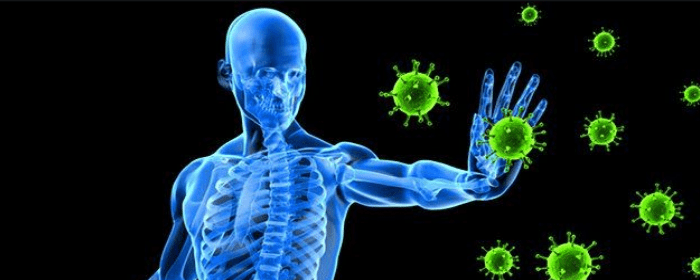
The human immune system can be one of our biggest assets or
one of our greatest foes. The immune system protects us against foreign
invaders like viruses and bacteria. It is essential for helping us maintain
immunity over a lifetime, whether from immunizations or previous infections. We
could not live without our immune systems.
On the other hand, the human immune system is the cause of
numerous diseases. Autoimmune
diseases like multiple sclerosis, ulcerative colitis, systemic lupus
erythematosus, and Crohn’s disease are caused by an immune system that
mistakenly attacks our own tissue. Organ and bone marrow transplants fail
because the body’s immune system rejects the transplant. When the immune system
is functioning normally, it is life-sustaining; however, when the immune system
falters, it can cause serious disease, suffering, and even death.
Compared to other diseases, the treatments for autoimmune
diseases and other diseases that involve the immune system are limited. Doctors
can prescribe steroids to knock down the immune response. These powerful drugs
can control disease flareups, but they aren’t a cure. Moreover, steroids cause
terrible side effects when taken long-term.
While there have been some recent developments in the treatment of certain autoimmune diseases (e.g. disease-modifying drugs for inflammatory bowel disease), medications are still limited. That is why scientists are actively studying the immune-modulating power of mesenchymal stem cells.
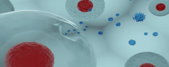
Mesenchymal stem cells exert a number of beneficial effects on the cells of the immune system. Mesenchymal stem cells can suppress T-cells, B-cells, dendritic cells, and natural killer cells (cells that participate in autoimmune diseases). Likewise, mesenchymal stem cells induce and affect the action of regulatory T-cells. This can help fine-tune the immune system, potentially shifting the balance from harmful to helpful immune system function.
Mesenchymal stem cells have been shown to be effective in various Phase I and Phase II clinical trials to treat multiple sclerosis, Crohn’s disease, lupus, ulcerative colitis, and even diabetes. While the clinical trials are often small—15-40 patients—the effects are impressive. Furthermore, treatment with mesenchymal stem cells is consistently safe; in study after study, the risk of serious adverse events is vanishingly small.
As with most fields of medicine, these clinical trials will need to be replicated in larger, Phase III trials. That being said, some large trials have already been conducted with favorable results. Perhaps the best example of a large trial testing the effect of mesenchymal stem cells on immune system function is in the field of transplantation medicine. The prestigious Journal of the American Medical Association (JAMA) published a clinical trial of 159 patients undergoing kidney transplants. Stem cell treatment reduced the incidence of kidney rejection, decreased the risk of opportunistic infection, and was associated with better kidney function 1 year after treatment.
The results from dozens of clinical trials suggest mesenchymal
stem cells are powerful modulators of immune cell function and have the
potential to one day be tools to treat diseases caused by the immune system. We
anxiously await further results from large, Phase III trials.
Reference: Gao, F., et al. (2016). Mesenchymal stem cells and immunomodulation: current status and future prospects. Cell Death & Disease. 2016, Jan; 7(1): e2062.
by admin | Mar 19, 2020 | Multiple Sclerosis, Mesenchymal Stem Cells
Multiple sclerosis is a chronic neurological disease that
affects the brain and spinal cord. In multiple sclerosis, an immune reaction
breaks down the covering around neuronal axons, myelin. Depending on where in
the brain or spinal cord this inflammation occurs, patients with multiple
sclerosis may experience weakness, a lack of sensation, double vision, difficulty
walking, difficulty with balance, problems with urination, dizziness, and/or
pain. Over time, patients with multiple sclerosis require assistive devices
such as canes or wheelchairs to get around, and some ultimately become
bedridden.
Multiple sclerosis can be divided into 4 types:
Clinically isolated syndrome – The first episode of MS; about two-thirds of people with a clinically isolated syndrome of MS will go on to have one of the other types listed below
Relapsing-remitting – Patients have symptoms for a
time, which resolve, but then eventually return
Secondary progressive – After some cycles of flare-ups
and remissions, the disease is present all the time; some patients with
relapsing-remitting disease develop secondary progressive MS
Primary progressive – Once symptoms start, they do
not resolve but instead get progressively worse
Fortunately, there are some treatments that can change the
course of multiple
sclerosis. There are several disease-modifying agents available for people
with relapsing-remitting disease and a few for progressive multiple sclerosis.
These drugs can reduce the rate at which symptoms get worse or extend the
length of time between flare-ups. Unfortunately, not everyone can tolerate
these drugs, and, in others, the drugs are not very effective. None of these
drugs is a cure for multiple sclerosis.
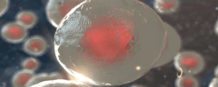
Researchers have turned to mesenchymal
stem cells as a possible treatment for multiple sclerosis. These cells have
the ability to regulate the immune system and support the nervous system. There
have been so many clinical trials of stem cells in multiple sclerosis that
researchers can now perform a meta-analysis on them. A meta-analysis is a
special study in which all trials on a particular topic are combined and
analyzed, i.e., a study of studies.
In their recent meta-analysis, Dr. Zhou and co-authors identified 9 clinical studies using mesenchymal stem cells to treat multiple sclerosis. They found that the rate of disease progression with stem cell treatment was 16% at 6 months and 35% at 1 year. This is a faster rate of decline than disease-modifying treatment, but better than no treatment at all. Importantly, the MS patients treated with stem cells had much more severe disease than average—it was a group for whom disease-modifying treatment had failed. From this perspective, stem cell treatment for MS was a great success.
Most patients had no evidence of disease activity at 6
months (72%) and 1 year (62%) after autologous
mesenchymal stem cell treatment. This a substantial duration of time to be
disease-free.
The authors noted that stem cells transplanted via the intrathecal
route (i.e. directly in the cerebrospinal fluid) resulted in a slower
progression of disability.
Taken together, these results are encouraging. The authors also concluded that mesenchymal stem cell treatment was safe. More work with larger patient groups are needed, but this is an exciting avenue of research.
Reference: Zhou, Y., et al. (2019). Autologous Mesenchymal
Stem Cell Transplantation in Multiple Sclerosis: A Meta-Analysis. Stem Cells
International. 2019, Volume 2019 |Article ID 8536785.
by admin | Feb 12, 2020 | Crohn's Disease, Mesenchymal Stem Cells
A recent study has shown how stem cells may be able to help Crohn’s disease patients who suffer from perianal fistulas. The researchers specifically investigated how stem cell therapy for Crohn’s Disease compared to conventional approaches including antibiotics and immunosuppressors. Because research is still in its infancy, there has been great interest in how to best address perianal fistulas in Crohn’s disease patients.
For their study, the scientists studied adults between the ages of 19 and 68. The patients were divided into three groups: one group received mesenchymal stem cell therapy applied locally, another received a combination of cellular and anti-cytokine therapy, and a third received a combination of immunosuppressors and antibiotics. The researchers looked at the impact of these three therapeutic interventions on the frequency of relapses of perianal fistulas and evaluated patients with the index of perianal activity of Crohn’s disease (PCDAI).
Their results showed that the combined cellular and anti-cytokine therapy improved perianal lesions in Crohn’s disease patients more so than did the immunosuppressor and antibiotic combination. Specifically, with the stem cell and anti-cytokine approach, fistulas remained closed longer and fistulas recurred less frequently. Future research should help to determine if and how this stem cell approach can provide an effective and safe long-term therapy for Crohn’s disease patients with perianal fistulas.
Reference: Knyazev, OV et al. (2018). Stem cell therapy for perianal Crohn’s disease. Ter Arkh, 90(3), 60-66.
by admin | Jan 15, 2020 | Exosomes, Kidney Disease, Mesenchymal Stem Cells, Stem Cell Research
Kidney diseases are among the most expensive and most debilitating diseases. Total costs are in excess of $50 billion a year, with $30 billion spent on people with end-stage renal disease including hemodialysis and kidney transplantation. People with kidney diseases have diminished quality of life, and substantial amounts of their time are devoted to medical treatment. Not surprisingly, researchers are aggressively pursuing novel therapies to treat kidney diseases before they result in end-stage renal disease. Stem cells and exosomes are among the most exciting and the most promising research topics in this area.
Most cells release tiny packets called extracellular
vesicles. The most notable extracellular vesicles are exosomes. While small,
exosomes are filled with high concentrations of potentially helpful substances
such as RNA, DNA, and proteins. While most cells release exosomes, researchers
are particularly interested in exosomes released by stem cells. It is within
these exosomes that stem cells pass along the substances that make stem cells
helpful in tissue repair and regeneration.
Zhang and
coauthors reviewed the recent advances that have been made using exosomes
to treat kidney diseases. Most of the work has focused on acute kidney injury
or AKI. Acute kidney injury can lead to
chronic kidney disease and kidney failure. Thus, if one could stop AKI, they
could potentially reduce the risk of chronic kidney disease.
Many different research groups have shown the power of
exosomes and other extracellular vesicles in treating acute kidney injury.
Exosomes taken from mesenchymal stem cells protected kidney cells from cell
death and fibrosis and helped them repair themselves. The
same was true of exosomes derived human umbilical cord stem cells.
Even stem cells taken from human liver cells were
able to improve kidney function after injury. There are many other examples.
Gatti
et al. reported that extracellular vesicles derived from human adult
mesenchymal stem cells could protect against acute kidney injury, but, most
impressively, also halted the progression of AKI to chronic kidney disease.
This finding has important implications for people who suffer from serious
acute kidney illnesses, such as kidney ischemia. It means that—if confirmed in
human studies—stem cell-derived extracellular vesicles can help treat kidney
disease in the short term and reduce the risk of that illness becoming a
chronic, debilitating problem.
Further research is needed in this field but, initial
results confirmed by many laboratories have created well-founded enthusiasm for
the future.
Reference: Zhang, W. et al. (2016). Extracellular vesicles
in diagnosis and therapy of kidney diseases. American Journal of Physiology – Renal Physiology. 2016, Nov 1;
311(5): F844-F851.
by admin | Jan 10, 2020 | Chronic Pain, Mesenchymal Stem Cells, Stem Cell Research
Chronic pain, that is, pain lasting for more than 12 weeks, is extremely common. As many as 1 in 3 Americans struggle with chronic pain, making it perhaps the most common physical condition that afflicts humans. Chronic pain is a source of significant suffering; it makes daily tasks more difficult, limits people’s ability to do the things they enjoy, and greatly diminishes the quality of life.
Perhaps the most frustrating aspect of chronic pain is that
there are usually no good treatments. Because the pain is chronic, patients who
try to treat chronic pain with medications must take drugs every day.
Acetaminophen and non-steroidal anti-inflammatories (NSAIDs) like ibuprofen can
help take away some suffering, but they are usually only partially effective.
Opioids are stronger, of course, but have such a high risk of causing
dependence and addiction that few doctors will prescribe for chronic non-cancer
pain. Physical therapy helps some, but not most people. Surgery may be able to
treat people with certain types of chronic musculoskeletal
pain; however, orthopedic surgery is a major ordeal with no guarantees of
success. Consequently, most people with chronic pain are left with very few
treatment options.
Perhaps this lack of chronic pain treatment options is what
makes recent stem cell research in chronic pain treatment so exciting.
Researchers are learning that mesenchymal
stem cells appear to be able to treat people with chronic pain. For
example, in a study of patients with low back pain who received treatment autologous
stem cells (their own stem cell populations purified, expanded, and
re-injected in their bodies), their pain and disability
decreased to the same levels as people who underwent major orthopedic
surgery (spinal fusion or total disc replacement). Another lab in Japan showed similar results.
The same benefit may occur in knee arthritis (osteoarthritis),
as well. Researchers showed that autologous stem cells were able to increase
the size of the knee
meniscus cartilage in a patient with severe knee arthritis. It should be
noted that damaged knee meniscus cartilage is one of the main sources of
chronic pain in knee osteoarthritis.
Of course, the number of patients treated in these studies is relatively small. Larger studies will be required to confirm that mesenchymal stem cells can treat chronic pain. However, these results are intriguing, since a staggering number of studies show autologous stem cell treatment to be safe. Indeed, mesenchymal stem cell treatment is now widely available in clinics and medical practices.
Reference: Waterman R. et al. (2011). Treating Chronic Pain with Mesenchymal Stem Cells: A Therapeutic Approach Worthy of Continued Investigation. Journal of Stem Cell Research & Therapy. 2011, S2 DOI: 10.4172/2157-7633.S2-001.
by admin | Dec 30, 2019 | Stem Cell Therapy, Adipose, Age Management, Mesenchymal Stem Cells, Stem Cell Research
Autologous stem cell treatments offer several advantages over other forms of stem cell treatment. In autologous stem cell treatment, a patient’s own stem cells are retrieved, processed, and injected back into the patient’s body. There is no need for a stem cell donor, and the entire procedure can take place in the same medical office. Since the patient’s own cells are used for an autologous stem cell treatment, there is no risk of disease transmission from a donor (because there is no donor) and no risk of rejection (because they are the patient’s own stem cells). Autologous stem cell treatments has some major benefits for the elderly.
Unfortunately, younger stem cells are better for
regenerative medicine than older stem cells are. Moreover, older people have
fewer stem cells that can be harvested than they did when they were younger. So
while autologous stem cell treatment is still advantageous, it becomes more difficult
to achieve as patients get older because their stem cells are fewer and less potent.
Making matters worse, older stem cells compete against more youthful stem
cells, making autologous stem cell treatments potentially even less effective
in older patients.
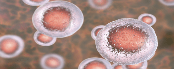
Fortunately, stem cell researchers are coming up with ways to make the most out of the stem cells that older patients still have. They still take a sample of tissue, such as fat, and harvest the stem cells contained within it. However, instead of injecting all stem cells from the sample (both older and youthful stem cells), researchers select and use only youthful stem cells. Furthermore, they make the treatments even more effective by injecting other substances (e.g. extracellular matrix) that help youthful stem cells survive, grow, and thrive.
To demonstrate the effectiveness of their approach,
researchers collected mesenchymal
stem cells from about a dozen older individuals aged 65 to 86 years old.
They then assorted the stem cells into different groups, separating youthful
from older stem cells. They then used special factors to help the youthful stem
cells grow, increasing the numbers by an impressive 17,000 times. So while only
8% of stem cells produced by older individuals are “youthful,” this laboratory
process increased those numbers to a point that they can be used for stem cell
treatments—even stored for future use!
The next phase of the research will be to inject these youthful stem cells into older patients and assess their effectiveness. However, even these preliminary results are exciting because they suggest that people of all ages can potentially benefit from autologous stem cell treatments, not just middle age and younger individuals.
Reference: Block, TJ et al. (2017). Restoring the quantity and quality of elderly human mesenchymal stem cells for autologous cell-based therapies. Stem Cell Research & Therapy. 2017 Oct 27;8(1):239.


 St. Petersburg, Florida