Human Umbilical Cord MSC-Derived Exosomes: Mapping the Future of Regenerative Medicine
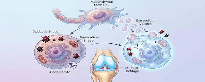
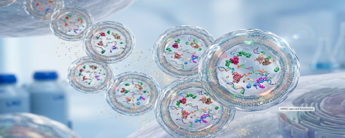
A Growing Area of Regenerative Medicine Human umbilical cord mesenchymal stem cell-derived exosomes, often abbreviated as hUCMSC-Exos, are becoming an increasingly important topic in regenerative medicine research. These tiny extracellular vesicles are released by human umbilical cord-derived mesenchymal stem cells and act as messengers between cells. Instead of using whole stem cells, exosome-based research focuses…