Living with a spinal cord injury changes how you move, feel, and function every day. You might be searching for more support in your recovery or looking into alternatives when other treatments have plateaued. At Stemedix, we provide access to regenerative medicine options, including stem cell therapy for spinal cord injury, designed to support your body’s healing potential. Our goal is to help you maintain independence and improve your quality of life through individualized care.
Stem cells for the treatment of spinal cord injury are being explored for their ability to support damaged nerve tissues and help reduce symptoms related to mobility, pain, and function. This therapy is not a cure, but it may serve as another layer of support in your recovery process. In this article, we will discuss how spinal cord injuries affect the body and how stem cell treatment may fit into your path forward.
Defining Spinal Cord Injury: Causes and Impact
A spinal cord injury doesn’t just affect mobility—it changes how the entire body communicates, functions, and adapts. Knowing how these injuries happen and what they cause can help you better plan your care and treatment options.
Common Causes of Spinal Cord Injury
A spinal cord injury is most often caused by sudden trauma or underlying medical conditions that disrupt nerve communication within the spine. These injuries commonly follow events such as vehicle crashes, major falls, sports-related impacts, or violent encounters.
Other cases develop from non-traumatic sources. These include conditions like spinal tumors, multiple sclerosis, and certain infections that interfere with the spinal cord’s structure and function. Degenerative diseases—such as spinal stenosis or arthritis—can also contribute to gradual nerve damage over time.
A spinal cord injury disrupts messages between the brain and the rest of the body. Where the injury occurs determines what parts of the body are affected. For example, if damage happens in the cervical spine, it can interfere with both arm and leg function. A lower injury in the lumbar region, by contrast, may impact only the hips and legs.
Immediate and Long-Term Effects on the Body
A spinal cord injury can result in paralysis, loss of sensation, and autonomic system dysfunction. Right after the injury, you might notice loss of movement, reduced feeling in certain areas, or changes in bladder and bowel control. These effects often appear quickly and may be temporary or permanent, depending on the severity.
As time passes, new challenges can appear. You may notice muscle weakness from disuse, skin breakdown from reduced movement, or respiratory changes if the injury is high enough to affect breathing muscles. Pressure injuries, also called pressure sores, and recurrent infections such as urinary tract infections are common secondary complications that require careful management. These long-term impacts highlight the importance of continuous support and well-structured care plans.
Classification of Spinal Cord Injuries by Severity and Location
Knowing where and how a spinal cord injury occurs helps you and your care team decide on the right approach to managing your recovery. The level and type of injury directly impact physical abilities, personal care needs, and long-term health planning.
Complete vs. Partial Injury Overview
A complete spinal cord injury causes total loss of motor and sensory function, while a partial injury retains some level of nerve signal transmission. If you’ve been diagnosed with a complete spinal cord injury, it means there’s no communication between the brain and the body below the injury site. This disconnect leads to full paralysis and loss of sensation below that point.
In contrast, partial, also called incomplete injuries, allow some signals to continue traveling along the spinal cord. You may notice that you still have some sensation, or you may be able to move certain muscles. These residual functions vary greatly between individuals. This classification matters because it plays a role in setting realistic goals for therapy and rehabilitation.
Differences Between Cervical, Thoracic, and Lumbar Injuries
The location of a spinal cord injury determines which parts of the body are affected. Cervical injuries often result in quadriplegia, thoracic injuries affect trunk and leg function, and lumbar injuries primarily impair lower limb control and bowel or bladder management.
Cervical injuries, those in the neck region, are often the most severe. They can impact movement and feeling in all four limbs, including breathing, swallowing, and arm function. These types are the most likely to require long-term assistive devices or full-time care.
Thoracic injuries occur in the middle section of the spine. While they typically spare arm movement, they may limit balance, torso strength, and control over abdominal muscles. It may be harder to sit upright or regulate body temperature below the injury level.
Lumbar injuries involve the lower spine and tend to affect the legs and lower body systems. Many people with lumbar-level injuries retain upper body function, but mobility challenges and changes in bladder or bowel function often follow. This type of injury may still allow for independent movement with the use of braces, walkers, or wheelchairs.
At Stemedix, we review all available medical records to understand your specific injury type and level before recommending any regenerative treatment option. This allows us to align our approach with your needs and current capabilities.
Stem Cell Therapy Explained: Purpose and Methods
Stem cell therapy for spinal cord injury involves introducing regenerative cells to promote repair and protect surviving tissue. These cells are introduced into areas near the injury site, where they may influence several healing processes. One of the primary actions is the regulation of the immune response, which helps reduce further damage caused by ongoing inflammation. In addition, stem cells may release biological signals that support the health of existing nerve cells and encourage the development of new connections within the nervous system.
Types of Stem Cells Used in Therapy
Stem cells for the treatment of spinal cord injury may sometimes include mesenchymal stem cells (MSCs), neural stem cells, and induced pluripotent stem cells (iPSCs). Each type works differently, but MSCs are the most frequently used in current therapeutic models. These cells are typically harvested from bone marrow or adipose (fat) tissue. They’re known for their ability to regulate inflammation and release molecules that promote healing.
Neural stem cells, on the other hand, are more specialized and are under investigation for their ability to integrate into damaged neural circuits. Induced pluripotent stem cells, adult cells reprogrammed into a more flexible, embryonic-like state, are still largely in the research phase. Although they offer broader potential, their use requires rigorous safety protocols to manage risks like tumor formation.
At Stemedix, we focus on therapies that use stem cells for the treatment of spinal cord injury with strong safety records and established handling procedures. Our team works closely with patients and referring physicians to coordinate care that is both informed by current science and centered on individual medical history.
Biological Actions of Stem Cells in Nerve Repair
Stem cells offer more than just cellular replacement—they create conditions in the body that support repair and healing. When applied to spinal cord injury, their effects can influence both immune activity and tissue regeneration.
Influence on Inflammation and Immune Response
Stem cells help regulate immune responses and reduce secondary damage from inflammation. After a spinal cord injury, inflammation can lead to further damage beyond the initial trauma. Immune cells flood the site, often destroying nearby healthy tissue in the process. This secondary damage can be just as limiting as the original injury.
Stem cells interact with this process by releasing bioactive molecules like cytokines and growth factors. These signals tell immune cells to calm their response and shift toward tissue support instead of attack.
This immune-modulating activity helps preserve nerve cells that might otherwise deteriorate. You’re not just adding cells—you’re also working with your body’s existing systems to limit further harm and stabilize the injury site.
Role in Regenerating Damaged Neural Tissue
Stem cell treatment for spinal cord injury may support the formation of new neural connections and repair mechanisms. Spinal cord damage disrupts the flow of signals between your brain and body.
To support repair, stem cells may promote three biological processes: axonal growth, remyelination, and cellular restoration. Axonal growth refers to the extension of nerve fibers that transmit signals. Without axons, communication between nerves stops.
Remyelination involves restoring the protective sheath around nerves, which allows electrical impulses to travel efficiently. In cases of spinal cord injury, this sheath often breaks down, leading to slower or blocked signals.
Studies show that certain types of stem cells, including induced pluripotent stem cells (iPSCs) and MSCs, can release growth factors that encourage axons to regrow and remyelinate existing nerves. These biological effects don’t occur all at once. They build over time as the cells interact with damaged tissue, guiding regeneration step by step.
At Stemedix, we focus on regenerative strategies that support your body’s efforts to recover. Stem cell therapy for spinal cord injury is structured to work with your body, using natural signaling processes to support healing at the cellular level.
Observed Outcomes from Stem Cell Treatments
Many individuals exploring regenerative options want to know what to expect from stem cell therapy. While results can differ, this section outlines some of the most reported effects based on real patient experiences and clinical data.
Enhancements in Mobility and Sensory Recovery
Some patients receiving stem cell treatment for spinal cord injury report improved strength, coordination, and sensation. These outcomes are often influenced by the level and completeness of the injury. For example, individuals with incomplete spinal cord injuries—where the spinal cord is damaged but not fully severed—have demonstrated positive changes in limb control, trunk stability, and tactile feedback following therapy.
Certain patients experienced measurable improvements in motor scores and sensory function within months after receiving stem cell injections. These functional changes, although not universal, suggest that the cells may support the body’s effort to reconnect or reinforce neural pathways.
The timing of intervention also plays a role. People who began stem cell treatment in the sub-acute phase (weeks after injury) have shown different patterns of recovery compared to those in chronic stages. It’s important to consider that early intervention may help maximize the biological environment for healing, but research is still ongoing to determine the full scope of response across timelines.
Reduction of Discomfort and Muscle-Related Symptoms
Stem cells have been observed to reduce spasticity and neuropathic pain associated with spinal cord injury. Spasticity, which causes involuntary muscle contractions, and nerve-related pain are among the most persistent challenges following spinal trauma. These symptoms can disrupt sleep, limit mobility, and interfere with rehabilitation.
Some patients who received mesenchymal stem cell (MSC) therapy reported decreased muscle stiffness and better pain control. Stem cell infusions modulated the immune response and contributed to reduced inflammation around damaged spinal segments. This shift may help explain why pain and tightness sometimes improve after treatment.
Relief from these symptoms can create opportunities for more active daily routines and improved engagement in physical therapy. While stem cell therapy is not a replacement for traditional pain management or rehabilitation, it may complement those approaches in supportive ways.
At Stemedix, we’ve seen that outcomes vary depending on the person’s overall health, injury characteristics, and treatment timing. Our role is to offer access to care designed around your condition while helping you understand how regenerative therapy might fit into your goals for living with a spinal cord injury.
The Treatment Process at Stemedix: Patient-Centered Approach
Every individual with a spinal cord injury presents a unique medical profile. At Stemedix, based in Saint Petersburg, FL, we align the treatment process with your personal health history and therapy goals to support your experience from evaluation through follow-up.
Importance of Diagnostic Information From Referring Physicians
Stemedix requires patients to provide medical imaging and records from their diagnosing physicians to determine eligibility for stem cell therapy. We rely on your existing records—such as MRIs, CT scans, and clinical summaries—to fully understand the scope of your spinal cord injury. This information gives us a starting point to evaluate whether stem cell therapy may be appropriate for your situation.
A detailed medical history helps our team determine the location and severity of your injury while also providing insight into how your body has responded to previous interventions. Accurate documentation from your physician allows us to move forward responsibly and reduce avoidable risks during the treatment process.
Tailoring Treatments to Individual Medical Histories
Each stem cell treatment for spinal cord injury is customized according to the patient’s health condition, injury level, and treatment goals. We look at a range of personal factors before planning treatment. These include the type of spinal cord injury you’ve experienced—whether complete or incomplete—as well as how long it has been since the initial trauma. Conditions like diabetes, autoimmune disorders, or chronic infections, as well as the medications you’re currently using, are all taken into account.
Administration Protocols and Safety Measures
Stemedix uses sterile, clinically guided protocols for administering stem cells. Each procedure is conducted in a controlled medical setting under the direction of trained clinicians. We use laboratory-tested biologics and sterile techniques to lower the risk of complications. All patients are closely observed before, during, and after the procedure.
Throughout treatment, we document patient responses, both for clinical records and to support communication with your existing care team. This consistent monitoring helps track progress and contributes to adjusting your care as needed over time. According to clinical studies, stem cell therapy has been associated with neurological improvements in some individuals with chronic spinal cord injuries, especially when introduced within a defined therapeutic window.
Patient Support Beyond Therapy
Recovery involves more than medical treatment alone. At Stemedix, we understand the physical and logistical challenges you may face when dealing with a spinal cord injury. That’s why we help coordinate accessible transportation and lodging for patients traveling from out of town, easing the burden of planning and focusing attention on your care.
To support your comfort during therapy, we provide access to mobility aids like wheelchairs and walkers, along with personal assistance when needed. Our team creates an accessible environment that allows you to move through treatment with as much comfort and independence as possible.
Start Your Recovery Journey with Stemedix Today
If you’re exploring advanced treatment options for spinal cord injury, our team at Stemedix is here to guide you every step of the way. We offer patient-focused care, treatment coordination, and support services designed around your individual needs. To learn more or speak with a care coordinator, call us at(727) 456-8968 or email yourjourney@stemedix.com.
Osteoarthritis and aging both impact your joints, but they are distinctly different processes. While natural aging causes gradual changes in joint structure, osteoarthritis is a diagnosed condition that results in progressive cartilage damage and joint pain. Recognizing the signs and symptoms of osteoarthritis, especially early osteoarthritis symptoms, is important for managing your joint health effectively.
At Stemedix, we focus on providing personalized stem cell therapy for osteoarthritis designed to support your body’s natural healing processes. This therapy is designed to support your body’s natural response to joint inflammation and help maintain joint function, helping you stay active and maintain your quality of life. Recognizing how aging and osteoarthritis differ allows you to make correct decisions about your treatment options. This article explains these differences and how stem cell therapy may play a role in your joint care journey.
Aging Joints vs. Osteoarthritis: What’s the Biological Difference?
You might notice your joints feel a bit stiffer or less flexible as you get older, but these changes don’t always mean you have a disease. Analyzing how normal aging differs from osteoarthritis can help you better manage your joint health.
Age-Related Joint Changes: Natural Degeneration Without Disease
Aging leads to gradual joint changes, even in healthy individuals. Over time, the cartilage that cushions your bones gradually loses water and becomes thinner. This reduces its ability to absorb shocks when you move. Additionally, the fluid that lubricates your joints may decrease, and your ligaments can become less flexible. These changes can lead to mild stiffness or discomfort, especially after periods of inactivity or overuse.
Despite these changes, natural aging does not usually cause inflammation or severe damage inside the joint. Most people with age-related joint changes continue their regular activities with only minor adjustments to how they move or exercise.
Osteoarthritis as a Diagnosed Condition
Osteoarthritis is a joint disease diagnosed by a healthcare professional, involving more than just natural wear and tear. In OA, the cartilage covering the bone ends breaks down at a faster rate, leading to direct bone-on-bone contact. This can cause inflammation, swelling, and damage to the tissues surrounding the joint.
Unlike simple aging, osteoarthritis leads to noticeable structural changes. You may find bone spurs forming and the joint lining thickening, which can reduce movement and increase pain. OA can develop in younger people, too, especially after injuries or if there is a family history of the condition.
Doctors use imaging tests, like X-rays or MRIs, along with physical exams and your medical history, to confirm osteoarthritis by identifying cartilage loss and narrowing of the joint space.
At Stemedix, we work with patients who have been diagnosed with osteoarthritis to explore treatment options that focus on supporting joint health and function. Understanding these differences helps you take the right steps toward managing your joint condition.
Signs and Symptoms of Osteoarthritis: What Goes Beyond Aging
You might notice similar patterns in how your joints feel as you get older, but osteoarthritis develops differently. The signs of this condition reflect disease, not just age.
Early Osteoarthritis Symptoms Often Overlooked
Early signs and symptoms of osteoarthritis often appear mild, so many people mistake them for normal aging. However, these early indicators are different in terms of both cause and progression. In healthy joints, occasional stiffness usually improves with light movement. With early osteoarthritis symptoms, there’s more happening beneath the surface.
You may feel a low level of inflammation around a joint, even though you haven’t had an injury. Mornings can start with stiffness that doesn’t ease after a few minutes. Some describe a subtle warmth or mild swelling around the joint, which may come and go. You might also hear or feel a soft grinding sound—known as crepitus—when moving the joint.
These early symptoms may not seem consistent or intense, which is why they’re easy to overlook. However, unlike age-related changes, early osteoarthritis symptoms often progress over time. The joint tissue continues to break down quietly, which makes it harder to manage later if ignored.
Later-Stage OA and Loss of Function
As osteoarthritis advances, the damage within the joint becomes more noticeable and harder to work around. Cartilage continues to erode, reducing your ability to move freely and without discomfort. At this stage, you may start walking differently without even realizing it. Some people adjust their posture or shift weight to avoid pain, which can affect the whole body.
The pain may no longer improve with rest. Even sitting still, the joint can throb or feel stiff. Everyday activities—like climbing stairs, driving, or exercising—may become more difficult.
Beyond discomfort, late-stage osteoarthritis can restrict how you live. It may affect work or limit how active you can be with friends and family. These limitations often stem from changes that are visible on imaging: narrowed joint spaces, worn cartilage, and bony growths.
At Stemedix, we support individuals who have already been diagnosed with osteoarthritis. If your symptoms are progressing or if early signs have been confirmed through evaluation, regenerative therapy options may be worth exploring with your care team.
Stem Cell Therapy for Osteoarthritis: How It Supports Joint Health
If you’ve been diagnosed with osteoarthritis, you may already be exploring ways to support your joints without adding more medications or surgeries to your treatment path. Stem cell therapy uses your body’s own resources to target joint changes at the cellular level.
The Role of Mesenchymal Stem Cells (MSCs)
Mesenchymal stem cells (MSCs) play a supportive role in joints affected by osteoarthritis. These adult stem cells are typically collected from your own fat tissue or bone marrow. In stem cell therapy for osteoarthritis, they are introduced into the area of joint damage, not to rebuild cartilage directly, but to interact with surrounding tissue in a meaningful way.
MSCs are known for their ability to send out helpful signals. Once in the joint, they influence nearby cells by releasing molecules that help reduce inflammation and support tissue maintenance. This type of signaling helps create a more balanced environment in joints where inflammation and cartilage breakdown are active. It’s not about forcing the body to regenerate but instead giving it tools to support itself.
Many patients come to us after their joints have become less responsive to conventional therapies. MSCs are being studied for how they influence pain levels, stiffness, and daily function over time. This therapy is part of an investigational field, and we guide each patient based on individual clinical history and medical documentation.
The Role of Chondrocytes in OA Treatment
Chondrocytes are the only cells found in healthy cartilage, and they’re responsible for producing and maintaining that cartilage. These cells don’t just sit in the joint; they actively respond to wear, damage, and changes in joint stress. Their presence is what keeps cartilage flexible and functional.
Research into regenerative medicine has started to examine how chondrocytes might be used in conjunction with stem cell strategies. Although this is still developing, scientists are looking at how these cartilage-producing cells may play a role in long-term joint support, especially in cases where cartilage breakdown is advanced. At this stage, these studies are helping the field better understand the cellular makeup of joint tissue and how it may respond to future therapies.
At Stemedix, we continue to follow the developments in the research closely to help our patients stay informed about the evolving landscape of regenerative care.
Evaluating Candidacy: What Patients Should Know at Stemedix
Before moving forward with stem cell therapy for osteoarthritis, it’s important to confirm that the condition has already been diagnosed. We focus on building treatments for those who already have clear documentation of their diagnosis.
The Importance of a Confirmed Diagnosis Before Treatment
At Stemedix, we work with individuals who have already received a confirmed diagnosis of osteoarthritis. Before starting therapy, we ask that you provide your existing medical records, including documentation such as imaging reports, physician notes, or clinical evaluations.
This information helps us design a treatment plan that’s based on what your care team has already identified. By reviewing accurate, up-to-date findings from your healthcare providers, we can approach your case with clarity and focus. Our role is to support your goals through regenerative therapy, not to replace the care already being provided by your doctor or specialist. We build on the foundation you already have in place, using that as a guide for what may come next.
How Treatment Plans Are Developed at Stemedix
Once you provide your records, our team reviews them carefully to determine if you are a fit for therapy. We look at your history, your imaging, and the specifics of your diagnosis. If we find that stem cell therapy for osteoarthritis may be appropriate for your situation, we will then create a plan tailored to your joint condition.
This is not a template approach. Every person’s joint health is different, and your treatment reflects that. It’s also important to know that our role is specific: we are not here to take over your full care. We support one part of your health journey while your main doctors continue to guide the rest.
What Makes Regenerative Medicine a Consideration for Osteoarthritis
Some individuals diagnosed with osteoarthritis are now exploring regenerative medicine as part of their symptom management plan. This approach is considered by those seeking alternatives that don’t involve major surgery or daily medication adjustments.
Investigational Status and Responsible Expectations
Stem cell therapy for osteoarthritis is currently categorized as an investigational procedure, and results can vary from one person to another. Some patients report improvements in joint mobility or reduced daily discomfort, but no outcome can be promised.
Stem cell therapy uses cells—often mesenchymal stem cells—that interact with the joint environment. These cells have been studied for their ability to release signals that may influence inflammation and tissue behavior. Current research focuses on how these signals might affect joint structures, such as cartilage and synovial tissue, in the context of chronic joint conditions like osteoarthritis.
You should approach this treatment with clarity and the understanding that it supports ongoing research. Your goals should be based on your current joint function, lifestyle, and medical history, not assumptions about universal outcomes.
Supporting Quality of Life Through Non-Invasive Approaches
Many individuals turn to stem cell therapy for osteoarthritis because it doesn’t involve major surgery or require significant downtime. This makes it a choice for those who are trying to maintain their daily routines or delay more invasive options.
If you’ve already tried physical therapy, exercise plans, or other forms of symptom management, you may be looking for additional support. This therapy may offer a path forward without disrupting what’s already working for you. Some patients use it alongside their existing care, not in place of it.
At Stemedix, we offer stem cell therapy to individuals who have already received a diagnosis of osteoarthritis. We work directly with each patient’s existing records and imaging to customize a treatment plan built around their condition and activity goals.
Staying Active While Managing OA
You don’t have to give up movement because of osteoarthritis. Small changes to your daily habits can help reduce strain on your joints and help you keep doing the things you enjoy.
Strategies Beyond Therapy: Daily Joint Care
Taking care of your joints everyday matters. Many people benefit from steady, low-impact movement such as walking, swimming, or cycling. These activities support strength and circulation without putting extra pressure on sensitive areas.
Your choice of footwear also plays a key role. Shoes with proper support help distribute your body weight evenly, which may reduce stress on your knees, hips, and ankles. If you’re walking for long periods or walking on uneven surfaces, braces or walking aids can help you stay steady and move more comfortably.
These tools and habits work alongside other treatment approaches. They won’t replace therapies, but they can support your mobility and independence over time.
Monitoring Progress Over Time
After you begin any treatment plan for osteoarthritis, it’s important to stay connected with your primary care provider. Regular check-ins allow your doctor to evaluate how your joints are responding over time and decide whether anything needs to be adjusted.
Tools like X-rays or MRIs can give more details about cartilage condition, joint space, or inflammation. If something changes, your doctor can catch it early and suggest the next steps. Staying involved in your care helps keep your progress on track and focused on your goals.
At Stemedix, we encourage every patient to stay active and work closely with their physician to manage their diagnosed osteoarthritis. Our role is to support you with treatment options that fit your condition, not to replace the care of your primary doctor.
Stemedix: Your Next Step Toward Joint Wellness
Osteoarthritis is more than just joint pain—it’s a condition that changes how your body moves and how you feel day to day. Recognizing the difference between normal aging and a diagnosed disease helps you decide what kind of care is right for you. If you’ve already been diagnosed, stem cell therapy for osteoarthritis may offer additional support for your current care plan. At Stemedix, based in Saint Petersburg, FL, we work with patients who are ready to take the next step with non-surgical options built around their existing records and goals. To speak with a Stemedix team member about stem cell therapy for osteoarthritis, call (727) 456-8968 or email us at yourjourney@stemedix.com. We’ll review your records and help you explore whether this therapy fits your joint care path.
Osteoarthritis (OA) is a common and painful joint condition that affects millions of people, causing significant discomfort and limiting mobility. As the cartilage in your joints breaks down over time, you may experience pain, stiffness, and swelling—common signs and symptoms of osteoarthritis. Recognizing early osteoarthritis symptoms can help you seek treatment sooner, potentially slowing the disease’s progression. While traditional treatments like pain medications and joint replacements have been the go-to options for many years, stem cell therapy for osteoarthritis is emerging as a promising alternative.
At Stemedix, we specialize in providing innovative regenerative medicine solutions to help you manage your osteoarthritis symptoms. This blog will guide you through the process of diagnosis, treatment, and recovery with stem cell therapy, showing you how this advanced approach can help heal your joints and improve your quality of life. Let’s explore how stem cell therapy for osteoarthritis could be the solution you’ve been searching for.
Step 1: Diagnosing Osteoarthritis
A successful approach to treating osteoarthritis (OA) begins with a confirmed diagnosis from your orthopedic doctor or primary care provider. If you suspect OA, it’s important to consult with a licensed physician who can assess your symptoms, review your medical history, and determine the severity of the condition.
Diagnosis is typically confirmed through a combination of physical examination and imaging studies, such as X-rays or MRIs. These tests help identify joint degeneration, cartilage loss, and other key signs of OA. Once your diagnosis is established, you can share your medical records and imaging results with the Stemedix team, and we will use this information to customize a regenerative medicine treatment plan tailored to your condition.
Signs and Symptoms of Osteoarthritis
Recognizing the signs and symptoms of osteoarthritis early on can make a significant difference in your treatment options. Common symptoms include joint pain, stiffness, and swelling, especially after activity or prolonged periods of rest. As the disease progresses, these symptoms may worsen, and you might start noticing a limited range of motion in your affected joints. By identifying early osteoarthritis symptoms, you can seek medical attention before the disease advances too far.
At Stemedix, we understand the importance of early detection and timely intervention. Our approach combines a thorough evaluation with advanced regenerative medicine treatments, like stem cell therapy, to help you address osteoarthritis early on and improve your quality of life.
Step 2: Understanding Stem Cell Therapy
Stem cell therapy is becoming an innovative and effective option for managing osteoarthritis, going far beyond temporary symptom relief. This approach utilizes mesenchymal stem cells (MSCs), which are harvested from sources such as adipose (fat) tissue or bone marrow. These powerful cells can differentiate into various cell types, including chondrocytes—specialized cartilage cells responsible for producing and maintaining healthy joint cartilage.
Chondrocytes are gaining attention in the field of regenerative medicine as a critical component in cartilage regeneration. As research continues to evolve, the role of chondrocytes in joint healing is proving to be especially promising for osteoarthritis treatment.
One of the standout benefits of stem cell therapy is its regenerative capacity. Rather than just masking discomfort, this therapy aims to restore damaged cartilage, improve joint function, and offer long-term relief. By stimulating the body’s natural healing response, stem cell therapy may help slow the progression of osteoarthritis and enhance mobility, allowing patients to return to the activities they enjoy.
How Stem Cells Help
Cartilage Regeneration: Stem cells support the repair of damaged cartilage by encouraging the formation of chondrocytes, which help rebuild and maintain the smooth, protective layer over joints.
Pain Relief: MSCs also possess anti-inflammatory properties, which can significantly reduce joint pain and swelling, leading to greater daily comfort.
Increased Mobility: With improved cartilage health and reduced inflammation, many patients experience better joint movement, flexibility, and overall functionality.
Stem cell therapy represents a breakthrough in how osteoarthritis is managed—focusing on healing from within rather than just managing symptoms. At Stemedix, our regenerative approach is designed to help you regain mobility, reduce discomfort, and experience a higher quality of life.
Step 3: Consultation with a Specialist
Before considering stem cell therapy for osteoarthritis, it’s essential to consult with a physician specializing in regenerative medicine. This consultation helps determine if stem cell therapy is the right treatment for your specific condition. Your physician will review your medical history, focusing on your symptoms and previous treatments to understand how OA has progressed and identify factors that may affect treatment options.
During the consultation process, patients are asked to provide existing medical documentation, including imaging results such as X-rays or MRI scans, along with a record of their diagnosis from their primary care provider or specialist. This information allows the Stemedix team to evaluate the extent of joint damage and determine whether stem cell therapy may be a suitable treatment option. Based on this review, a customized regenerative medicine plan is created to support each patient’s specific condition and goals.
What to Expect During the Consultation
Medical History Review: The physician will ask detailed questions about your symptoms, such as when the pain started, how it has progressed, and what treatments have been tried.
Test Results: Imaging results provide a clear picture of joint health. Your doctor will review these images to evaluate the extent of cartilage damage and how advanced the osteoarthritis is. This will help to decide whether stem cell therapy can be a suitable solution or if other treatments might be required.
Treatment Plan: Once the physician has all the necessary information, they will discuss the potential benefits and risks of stem cell therapy. You will learn about the process, from how stem cells are processed and injected into the affected joint to the expected timeline for recovery and improvement. The physician will outline the steps involved in the procedure and answer any questions you may have, ensuring you have a clear understanding of what to expect.
At Stemedix, we believe in making sure you feel fully informed and comfortable with every step of the process. Our team is here to guide you through your journey, helping you make the best decision for your joint health and overall well-being.
Step 4: The Stem Cell Therapy Procedure
Stem cell therapy for osteoarthritis is a minimally invasive procedure that can provide a less disruptive alternative to traditional treatments like surgery. The process involves harvesting stem cells from your own body, typically from bone marrow or adipose tissue (fat), which are then processed and injected directly into the affected joint to stimulate healing.
The procedure is generally well-tolerated and performed under local anesthesia, meaning you’ll remain awake but pain-free during the process. The injection itself is relatively quick, and the recovery time is typically short. Most patients can resume their normal daily activities within just a few days. However, it’s important to avoid high-impact or strenuous activities during the early stages of healing to allow the tissue to regenerate properly.
What Happens During the Procedure?
Stem Cell Harvesting: To begin, a small sample of either bone marrow or fat tissue is collected from your body. This is usually done from the hip or abdomen, areas where these tissues are readily accessible. The process is minimally invasive, requiring only a small incision or needle insertion.
Stem Cell Processing: Once the tissue is harvested, it’s processed in a laboratory setting to isolate the stem cells. The stem cells are then prepared for injection into the damaged joint. This step guarantees that only the necessary cells are used to promote healing.
Injection into the Joint: After processing, the stem cells are carefully injected into the affected joint, where they begin to work on repairing damaged cartilage, reducing inflammation, and promoting overall joint regeneration. This targeted injection allows the stem cells to focus their healing efforts directly where they are needed most.
At Stemedix, we pride ourselves on using advanced techniques and providing clear instructions throughout the procedure. Our medical team makes sure that you are informed and comfortable at every stage of the therapy, helping you feel confident as you take steps toward managing your osteoarthritis more effectively.
Step 5: Recovery and Long-Term Results
After undergoing stem cell therapy for osteoarthritis, the recovery process is vital for ensuring the best results. This stage involves a period of rest and limited activity to give the stem cells time to take effect and begin regenerating the damaged tissue. While the procedure is minimally invasive, your body still needs time to heal and respond to the therapy.
In the weeks following the procedure, you will likely notice gradual improvements. These include reduced pain, enhanced mobility, and better overall joint function. However, it’s important to understand that the full benefits of stem cell therapy can take several months to become fully apparent, as the stem cells work overtime to regenerate cartilage and restore joint health.
Post-Procedure Care
Rest: In the immediate weeks after the procedure, it’s recommended to limit strenuous activities. Resting and avoiding high-impact exercises during this period will allow the stem cells to do their work without disruption.
Follow-Up Appointments: Regular follow-up visits with your physician are essential to track the progress of the therapy and evaluate how well the joint is healing. These appointments will help identify any adjustments needed to optimize your recovery and provide the best possible outcome.
Physical Therapy: Engaging in physical therapy after stem cell therapy is highly beneficial. Physical therapy focuses on improving mobility, strengthening the muscles surrounding the joint, and preventing stiffness. By working with a qualified therapist, you can further enhance the healing process and regain function more effectively.
At Stemedix, based in Saint Petersburg, FL, we take an extensive approach to care, guiding you through each phase of the recovery process. Your dedicated care coordinator will help you stay on track, offering support and resources to maximize the success of your treatment. With patience and proper care, stem cell therapy can provide lasting relief from the symptoms of osteoarthritis, improving your overall quality of life.
Step 6: Maintaining Joint Health
After experiencing the benefits of stem cell therapy for osteoarthritis, it’s essential to adopt a proactive approach to maintaining your joint health. The therapy may help regenerate cartilage and reduce pain, but long-term success depends on how you care for your joint moving forward. Incorporating specific lifestyle changes, ongoing physical therapy, and healthy habits will help support the healing process and prevent further degeneration of the joint.
Tips for Long-Term Joint Health
Low-Impact Exercise: Regular, low-impact exercise is one of the best ways to maintain joint function and mobility. Activities like swimming, walking, or cycling can help keep your joints flexible without putting undue stress on them. These exercises promote circulation and strengthen the muscles surrounding the joint, which helps support it during movement.
Anti-Inflammatory Diet: Nutrition plays a key role in joint health. Consuming foods that are rich in omega-3 fatty acids, antioxidants, and vitamin D can reduce inflammation in the body. This helps keep the joints from becoming inflamed, which can lead to additional damage over time.
Physical Therapy: Continuing with physical therapy can be highly beneficial, even after the stem cell procedure. Regular sessions can enhance the strength and flexibility of the muscles surrounding your joint, which helps reduce the risk of further damage. Additionally, physical therapy can improve your range of motion, making movement easier and more comfortable.
At Stemedix, we encourage patients to take an active role in their joint health after treatment. By following these steps and working closely with your care coordinator and medical professionals, you can maintain the progress made from stem cell therapy and prevent future osteoarthritis complications.
Trust Stemedix for a New Path to Joint Health
Recognizing the signs and symptoms of osteoarthritis early is essential for managing the condition effectively. If you’re experiencing early osteoarthritis symptoms, such as joint pain, stiffness, or swelling, it’s important to seek a professional evaluation as soon as possible. Early diagnosis and intervention can help slow the progression of the disease and improve your quality of life. At Stemedix, we offer advanced stem cell therapy to regenerate damaged cartilage and restore joint function, providing long-term relief from osteoarthritis.If you’re ready to explore how stem cell therapy can help you manage osteoarthritis, contact Stemedix today. Our team of experts is here to guide you through every step of the process and guarantee the best possible outcomes. Call us at (727) 456-8968 or email us at yourjourney@stemedix.com to schedule your consultation and take the main step toward healing.
Osteoarthritis can make everyday movements difficult, limiting mobility and causing persistent joint pain. If you have been relying on pain medications or considering surgery, there is another option. At Stemedix, we offer regenerative medicine treatments that address joint damage at the source, helping you regain function without invasive procedures.
Through therapies like stem cell and platelet-rich plasma (PRP) injections, regenerative medicine promotes natural healing by stimulating tissue repair and reducing inflammation. Unlike traditional treatments that only manage symptoms, these therapies work to restore cartilage and improve joint function over time.
For those seeking regenerative medicine in Saint Petersburg, FL, Stemedix offers advanced, full-service, patient-focused care tailored to each individual’s needs. From airport and appointment transportation to providing wheelchairs, walkers, and shower chairs, Stemedix guarantees a comfortable and supported experience throughout your treatment journey. Our team of experts develops personalized treatment plans designed to help you stay active and avoid surgery.
Osteoarthritis and Its Impact on Joint Health
Osteoarthritis (OA) is a progressive joint disease that causes cartilage deterioration, leading to pain, stiffness, and mobility issues. It is the most common type of arthritis, affecting millions of people worldwide. It develops as the protective cartilage that cushions the ends of bones begins to wear down. Over time, this deterioration leads to increased friction between bones, causing pain, stiffness, and inflammation. The condition primarily affects joints that bear the most weight, such as the knees, hips, and spine. However, it can also impact the hands and other areas, making even simple movements uncomfortable.
Although aging plays a role in its progression, osteoarthritis is not just a condition that affects older adults. Joint injuries, repetitive stress, obesity, and genetic factors can contribute to early onset and severity. Many individuals begin to notice symptoms when stiffness becomes more persistent, and activities that were once effortless start to feel more challenging. Without intervention, the condition can continue to worsen, affecting overall mobility and quality of life.
How Osteoarthritis Affects Joint Function and Mobility
Joints rely on smooth cartilage surfaces for pain-free movement. As osteoarthritis progresses, cartilage breaks down, exposing the underlying bone. This increases friction during movement, triggering inflammation and pain. Swelling and stiffness can make it difficult to bend, straighten, or bear weight.
As osteoarthritis advances, bone spurs can form around the joint, restricting motion and adding to discomfort. Fluid accumulation within the joint capsule contributes to swelling, further limiting movement. In severe cases, the cartilage loss becomes so pronounced that bones grind directly against each other, resulting in chronic pain and mobility impairments.
The impact of osteoarthritis extends to daily activities. Tasks like walking, climbing stairs, or standing for long periods become increasingly difficult. Individuals may also experience weakness or instability, increasing the risk of falls or further injury, which can substantially affect independence and overall well-being.
For those struggling with osteoarthritis, finding effective treatment options is essential. At Stemedix, we provide regenerative medicine treatments designed to support natural joint repair, helping individuals regain function and reduce pain without relying solely on medications or surgery.
The Role of Regenerative Medicine in Osteoarthritis Treatment
Regenerative medicine is an approach that utilizes the body’s natural healing mechanisms to repair and restore damaged tissues. This includes stem cell therapy and specialty cells, which can replace and rebuild cartilage, reduce inflammation, and improve joint function, which help regenerate cartilage, reduce inflammation, and improve joint function.
How Stem Cell Therapy Can Aid in Joint Repair
Stem cell therapy introduces specialized cells into the affected joint to help repair and regenerate damaged tissue. These cells are typically sourced either from the patient’s own bone marrow or adipose (fat) tissue, or from carefully screened donor tissue, depending on the treatment approach. Once introduced into the joint, they interact with the surrounding environment to promote healing. Studies suggest that stem cells, especially chondrocytes (Cartilage Cells) and their respective exosomes can contribute to cartilage regeneration, slow the progression of osteoarthritis, and improve overall joint function.
One of the main benefits of stem cell therapy is its ability to address joint degeneration at a cellular level. Unlike pain medications or corticosteroid injections, which only provide temporary symptom relief, stem cells work to support the restoration of tissue over time. Many individuals who undergo stem cell therapy report gradual improvements in pain levels, joint flexibility, and overall mobility. While results vary, research continues to explore how stem cell treatments can enhance long-term joint health.
Platelet-rich plasma (PRP) Therapy: A Complementary Treatment
PRP therapy is another regenerative medicine treatment that can support joint healing. This therapy involves drawing a small sample of the patient’s blood, processing it to concentrate the platelets, and injecting the enriched plasma into the affected joint. Platelets contain growth factors that stimulate tissue repair, reduce inflammation, and enhance the body’s natural healing processes.
For osteoarthritis patients, PRP therapy can help manage pain and improve function by promoting cartilage repair. When used in combination with stem cell therapy, PRP may enhance the effects of treatment by creating an environment that supports regeneration. Some individuals experience noticeable improvements in joint comfort and mobility after a series of PRP injections, making it a valuable option for those seeking alternatives to surgery.
At Stemedix, we provide regenerative medicine treatments designed to support joint repair and improve quality of life. By offering stem cell therapy and PRP therapy, we help individuals take a proactive approach to osteoarthritis management, reducing pain and enhancing mobility without the need for invasive procedures.
Benefits of Regenerative Medicine for Osteoarthritis
Regenerative medicine treatments provide several benefits for osteoarthritis patients looking for a non-surgical solution:
Non-Surgical Treatment with Minimal Recovery Time
Regenerative medicine treatments offer a non-invasive approach to managing osteoarthritis. Unlike joint replacement surgery, which requires extensive incisions, hospital stays, and long recovery periods, regenerative therapies involve targeted injections. This means patients experience minimal discomfort during the procedure and can return to their normal activities much sooner. Most individuals can resume daily routines within days, making it a practical option for those who want to avoid the risks and downtime associated with surgery.
Because these treatments do not require general anesthesia or large incisions, the risk of complications is lower compared to surgical interventions. Many patients choose regenerative medicine to maintain an active lifestyle while managing osteoarthritis symptoms effectively.
Promoting Natural Healing and Tissue Regeneration
Traditional osteoarthritis treatments, such as pain medications and steroid injections, primarily focus on symptom relief. While they may reduce discomfort temporarily, they do not contribute to long-term joint health. Regenerative medicine takes a different approach by supporting the body’s natural ability to heal itself.
Stem cell therapy and PRP therapy introduce healing elements directly into the affected joint, encouraging the repair of damaged cartilage and tissues. Stem cells can develop into various types of cells needed for joint repair, while PRP provides growth factors that stimulate tissue regeneration. Over time, these therapies may help slow osteoarthritis progression, preserving joint function and mobility for longer.
Reducing Pain and Inflammation
Inflammation is one of the primary drivers of osteoarthritis pain. As the cartilage wears down and bones begin to rub together, the body responds with increased inflammation, leading to swelling, stiffness, and discomfort. Regenerative medicine treatments target this underlying inflammation rather than simply masking pain.
By introducing stem cells and PRP into the affected joint, these treatments help regulate the inflammatory response. This can lead to sustained pain relief and improved joint function over time. Many individuals report noticeable reductions in pain and stiffness, allowing them to engage in physical activities with greater ease.
At Stemedix, we specialize in regenerative medicine treatments that help individuals manage osteoarthritis without relying on invasive procedures. By offering innovative stem cell therapies that promote healing, reduce inflammation, and restore function, we provide a path to long-term joint health and improved mobility.
Is Regenerative Medicine the Right Treatment for You?
Regenerative medicine is an effective treatment option for individuals with mild to moderate osteoarthritis who are seeking a non-surgical alternative to managing their condition. If you have joint pain, stiffness, and decreased mobility that have not responded well to traditional treatments, regenerative therapies may be an excellent choice.
This approach is especially suited for people who wish to avoid the risks, recovery time, and complications associated with joint replacement surgery. If you are looking to regain function, reduce pain, and maintain mobility without the need for invasive procedures, regenerative medicine may be the solution for you.
What to Expect During the Treatment Process
The process for regenerative medicine treatments typically involves injecting stem cells, specialty cells, or even platelet-rich plasma into the affected joint. The procedure is minimally invasive, usually performed in an outpatient setting, and involves very little discomfort.
Before the injection, your healthcare provider will make sure that the area is numbed to minimize any discomfort during the procedure. The stem cells, or PRP, are then injected directly into the affected joint. While the treatment itself is relatively quick, it may take several weeks for noticeable improvements to appear. Over the following months, the benefits of the therapy continue to unfold, helping to promote tissue regeneration, reduce inflammation, and alleviate pain.
At Stemedix, we specialize in providing personalized regenerative medicine treatments tailored to your unique needs. If you’re considering this option for osteoarthritis, our experienced team can help guide you through the process, ensuring that you receive the best care and support throughout your treatment journey.
Regenerative Medicine vs. Traditional Joint Replacement Surgery
Joint replacement surgery becomes necessary when osteoarthritis has progressed to a point where the damage to the joint is severe and non-surgical treatments no longer provide relief. In these cases, the cartilage has deteriorated, causing bones to rub against each other, resulting in debilitating pain and limited movement. When conservative treatments like medication, physical therapy, and injections fail to alleviate symptoms, joint replacement may be the only remaining option. This procedure involves removing the damaged cartilage and bone and replacing it with an artificial joint.
However, joint replacement surgery comes with risks, including infection, blood clots, and a lengthy recovery period. For many patients, the idea of undergoing surgery and spending months recovering is stressful.
Advantages of Regenerative Medicine Over Surgery
Regenerative medicine offers a compelling alternative to joint replacement by addressing joint deterioration early in the process. By harnessing the body’s natural healing abilities, regenerative treatments like stem cell therapy and PRP (Platelet-Rich Plasma) therapy target the root causes of joint pain and inflammation.
One of the most notable benefits of regenerative medicine is the shorter recovery time compared to surgery. Since regenerative treatments involve injections rather than incisions, patients typically experience less pain and minimal downtime and can return to their daily activities much faster. Additionally, regenerative medicine carries fewer risks, as it’s a non-invasive procedure that doesn’t require general anesthesia or long hospital stays.
Another significant advantage is that regenerative treatments may help delay or even prevent the need for joint replacement altogether. By stimulating tissue regeneration, regenerative medicine can slow the progression of osteoarthritis and preserve joint function.
Cost Considerations: Regenerative Medicine vs. Joint Replacement
While joint replacement surgery can cost tens of thousands of dollars, regenerative medicine treatments tend to be more affordable. Surgery often requires extensive post-operative care, including physical therapy and follow-up visits, which can add up in costs. Additionally, joint replacement surgery typically involves a long recovery period, where additional medical expenses may arise.
On the other hand, regenerative medicine offers a cost-effective alternative. Since the procedures are minimally invasive, they generally don’t require the same level of post-treatment care and rehabilitation. By avoiding the need for surgery, patients can save money in the long term, while also potentially experiencing better outcomes without the extensive recovery times.
At Stemedix, we provide advanced regenerative medicine treatments tailored to your needs, offering an affordable, non-surgical alternative that may help you avoid joint replacement surgery. Our team of experienced providers is here to guide you through your treatment options and help you make the best decision for your joint health.
Stemedix: Your Partner in Regenerative Medicine for Osteoarthritis
At Stemedix, we specialize in offering advanced regenerative medicine in Saint Petersburg, FL, designed to provide relief from osteoarthritis and restore joint function. Our clinic is known for its commitment to providing the highest level of care, using innovative treatments like stem cell therapy, specialty cells, and PRP (plat-r-p) therapy. These treatments are tailored to help manage pain, reduce inflammation, and regenerate damaged tissues, ultimately improving mobility and quality of life.
We understand that each patient’s needs are unique, which is why we focus on personalized care and treatment options. Whether you’re looking to delay surgery or improve your joint function, Stemedix is dedicated to helping you achieve your goals with effective, non-surgical alternatives.
How Stemedix Tailors Treatments for Osteoarthritis Patients
At Stemedix, we take a personalized approach to every case of osteoarthritis. Our medical team works closely with each patient to design a customized treatment plan that suits their specific condition. We don’t believe in a generalized approach—our regenerative medicine treatments are based on evidence-based practices that are aimed at providing the most effective results for your joint health.
Whether it’s stem cell therapy to regenerate damaged cartilage or PRP therapy to reduce inflammation and promote healing, we combine these treatments to maximize joint restoration and pain relief. Our goal is to enhance your mobility and quality of life while avoiding the need for invasive surgeries.
Patient-Centered Care at Stemedix
At Stemedix, based in Saint Petersburg, FL, patient care is at the heart of everything we do. From the moment you walk through our doors, you’ll experience a welcoming and supportive environment. We understand that going through osteoarthritis treatment options can be overwhelming, which is why we assign dedicated care coordinators to guide you every step of the way.
Our care coordinators make sure that you have all the information you need to make well informed decisions about your treatment. They work closely with you to schedule appointments, answer questions, and monitor your progress throughout the healing process. We prioritize your comfort, convenience, and well-being, ensuring that your experience is smooth and stress-free, from consultation to recovery.
Explore Regenerative Medicine as a Viable Osteoarthritis Treatment Option at Stemedix
Regenerative medicine offers a valuable treatment option for individuals experiencing osteoarthritis in Saint Petersburg, FL. Unlike conventional methods that focus solely on symptom management, regenerative therapies such as stem cell therapy and PRP (Platelet-Rich Plasma) therapy work to restore joint health by promoting natural healing. These treatments support tissue repair, reduce inflammation, and encourage cartilage regeneration, which can enhance mobility and long-term joint function.
Stemedix specializes in regenerative medicine in Saint Petersburg, FL, providing a non-surgical alternative for those seeking relief from osteoarthritis. By leveraging innovative therapies, Stemedix helps patients regain mobility and improve their quality of life without the risks and extended recovery associated with surgery.
If you are looking for an advanced, non-invasive approach to managing osteoarthritis, Stemedix is here to help. Contact us today at (727) 456-8968 or email yourjourney@stemedix.com to learn more about how regenerative medicine can support your joint health.
Multiple sclerosis (MS) is a progressive neurological condition that affects millions of people worldwide. As this autoimmune disease disrupts the central nervous system, it leads to symptoms such as muscle weakness, numbness, and cognitive issues. In recent years, stem cell therapy has emerged as a promising treatment to alleviate these symptoms and potentially slow the progression of the disease.
At Stemedix, we recognize the challenges that MS patients face, particularly as the disease advances. We are dedicated to exploring stem cell treatments for multiple sclerosis as a potential solution. Stem cell therapy offers new hope by targeting the underlying causes of MS, especially the destruction of myelin—the protective sheath around nerve fibers. Myelin loss disrupts communication between the brain and the body, contributing to MS symptoms. Stem cells have the unique ability to regenerate damaged tissues, reduce inflammation, and modulate the immune system, which is critical in autoimmune diseases like MS.
Stem cell treatments for multiple sclerosis aim to restore function and slow the disease’s progression. Whether you’re experiencing early warning signs of multiple sclerosis, such as unexplained fatigue, numbness, or vision problems, or have been living with the disease for some time, stem cell therapy could offer a pathway to managing symptoms and improving your quality of life. In this article, we will explore how stem cell therapy for MS works, the scientific mechanisms behind it, and what you can expect from the treatment process. At Stemedix, we’re committed to helping you understand how stem cell treatments can make a difference in your journey with MS.
Stem Cell Therapy: A Game Changer for MS Treatment
Stem cell therapy offers a new approach to treating multiple sclerosis (MS), offering hope for many individuals living with this challenging condition. Understanding stem cells and their unique capabilities is essential in recognizing how stem cell therapy can be a powerful tool in MS treatment.
What Are Stem Cells?
Mesenchymal stem cells (MSC’s) are unique cells with the remarkable ability to transform into different cell types in the body. Known for their regenerative properties, they serve as the building blocks of life. In multiple sclerosis, stem cells can repair damaged tissues, including nerve cells affected by the disease. Unlike other cell types, stem cells are undifferentiated, meaning they can develop into specialized cells, such as those needed to regenerate the myelin sheath—the protective covering around nerve fibers often damaged in MS. While other treatments primarily manage symptoms or inflammation, stem cell therapy works to repair the underlying damage to the nervous system, making it a vital tool in regenerative medicine focused on healing rather than just symptom control.
Specialty Stem Cells in Multiple Sclerosis Treatment
Stem cell therapy for Multiple Sclerosis not only focuses on reducing inflammation but also on regenerating and repairing nerve damage. Certain specialized stem cells play an important role in this process:
Neural Stem Cells (NSCs): These cells have the potential to develop into various types of nerve cells, supporting the repair of damaged neurons and promoting neuroprotection. They may help restore function by replacing lost or injured nerve cells in MS patients.
Oligodendrocyte Precursor Cells (OPCs): Oligodendrocytes are responsible for producing myelin, the protective sheath around nerve fibers that is damaged in MS. Stem cell-derived OPCs aim to restore myelin, improving nerve function and slowing disease progression.
Schwann Cells: While primarily associated with the peripheral nervous system, Schwann cells play a role in myelin regeneration and nerve repair. Their regenerative properties make them an important consideration for supporting neural function in MS patients.
By incorporating these specialized stem cells into treatment strategies, regenerative medicine aims to go beyond symptom management and actively promote nerve repair and functional recovery. Stemedix continues to provide therapies informed by the latest research in stem cell applications for MS.
How Stem Cells Can Help MS Patients
Multiple sclerosis (MS) occurs when the immune system attacks the myelin, disrupting communication between the brain and the body. This leads to symptoms like numbness, muscle weakness, and cognitive challenges. Stem cells have the unique ability to regenerate the myelin sheath, repairing this damage. A key benefit of stem cells is their ability to reduce inflammation, which is central to the ongoing nerve damage in MS. By modulating the immune response, stem cells help control inflammation, providing symptom relief and potentially slowing disease progression. Stem cells may aid in regenerating damaged nerve cells and improving mobility, coordination, and cognitive function, making them a promising treatment option for MS.
At Stemedix, we recognize the challenges that come with MS, and we are committed to providing personalized stem cell treatments designed to address the root causes of the disease. Our goal is to offer a pathway to improved quality of life, aiming to slow the progression of MS and provide patients with the relief they need. If you’re considering stem cell therapy for MS, Stemedix is here to guide you every step of the way.
The Scientific Mechanisms Behind Stem Cell Treatments for MS
Stem cell therapy has become one of the most promising approaches to treating multiple sclerosis (MS). By targeting the underlying causes of the disease, stem cells offer a potential solution for repairing damage to the nervous system and improving overall function. Understanding the scientific mechanisms behind stem cell treatments can provide greater clarity on how these therapies work and why they hold so much potential for MS patients.
How Stem Cells Repair Damaged Myelin
Myelin is the protective covering around nerve fibers in the central nervous system, and its destruction is a key characteristic of multiple sclerosis (MS). When myelin is damaged, nerve signals cannot travel properly, resulting in symptoms like muscle weakness, numbness, and cognitive issues.
Stem cells can help regenerate myelin by transforming into oligodendrocyte precursor cells (OPCs), which produce new myelin. This regeneration improves nerve signal transmission and enhances overall function. Research, including animal models and early human trials, has shown promising results, with stem cell therapy leading to myelin repair and functional recovery. While still considered an emerging treatment, stem cell therapy’s potential to repair myelin offers hope for reducing MS symptoms and slowing disease progression.
Immune System Regulation
In multiple sclerosis, the immune system erroneously attacks myelin, causing progressive damage. Stem cells can modulate the immune system, reducing its overactive response and preventing further damage to the nervous system. This immune-modulating effect is critical in treating autoimmune conditions like MS.
Stem cells can reset the immune system by influencing T cells and B cells, which play a key role in attacking myelin. Ongoing research is investigating how stem cells can rebalance this immune response, potentially leading to long-term disease stabilization and fewer relapses. This immune modulation is a key mechanism of stem cell therapy for MS, addressing the disease’s root cause rather than merely managing its symptoms.
Reducing Inflammation and Enhancing Nerve Function
Chronic inflammation is another key feature of multiple sclerosis, contributing to the ongoing destruction of nerve cells and myelin. Stem cells can help combat this inflammation by producing anti-inflammatory cytokines, which are molecules that regulate the immune response. By reducing inflammation, stem cells help prevent further damage to the nervous system and support the body’s healing process.
Additionally, stem cells play a vital role in encouraging the repair of nerve cells and improving communication between the brain and the body. The regeneration of myelin and the reduction of inflammation work together to enhance nerve function, which can lead to improvements in mobility, coordination, cognitive function, and overall quality of life for MS patients.
Stem cell treatments for MS offer a multifaceted approach that addresses the damage caused by the disease, from repairing the myelin sheath to modulating the immune system and reducing inflammation. These scientific mechanisms provide a strong foundation for why stem cell therapy is considered a potential game-changer for those living with multiple sclerosis.
Types of Stem Cell Therapies for MS: Which One is Right for You?
Stem cell therapy is rapidly emerging as a viable option for individuals living with multiple sclerosis (MS). However, there are different types of stem cell therapies, each with unique processes and potential benefits. Understanding the different options available can help you make an informed decision about the treatment that’s best for you.
Autologous Stem Cell Therapy
Autologous stem cell therapy uses the patient’s own stem cells, offering a highly personalized treatment for multiple sclerosis (MS). The process begins with collecting stem cells from the patient’s bone marrow or blood. These cells are then purified in a laboratory and reintroduced into the body to help regenerate damaged tissues, repair myelin, and modulate the immune system.
A significant benefit of autologous stem cell therapy is the elimination of immune rejection, as the cells are derived from the patient’s own body. This reduces complications associated with foreign tissue. However, challenges include the time-consuming, expensive nature of the process and limited stem cell availability in some patients, especially older individuals. Despite these hurdles, it remains a popular and effective MS treatment.
Allogeneic Stem Cell Therapy
Allogeneic stem cell therapy uses stem cells from a healthy donor rather than the patient’s own cells. These donor cells are harvested, processed in a lab, and transplanted into the patient. This approach is helpful when a patient’s stem cells are not viable or when a quicker stem cell replenishment is needed.
One key benefit is the immediate availability of high-quality donor cells that can regenerate tissue, repair myelin, and modulate the immune response in MS patients.
Mesenchymal Stem Cells (MSCs)
Mesenchymal stem cells (MSCs), typically sourced from umbilical cord tissue (UCT), adipose tissue, or bone marrow, hold significant promise for treating multiple sclerosis (MS). These cells are known for reducing inflammation, promoting tissue repair, and aiding in the regeneration of damaged myelin. MSCs also modulate the immune system, addressing the autoimmune response driving MS progression.
MSC therapy has garnered attention for its potential to repair MS-related damage while addressing immune dysfunction. These cells release anti-inflammatory cytokines, alleviating chronic inflammation. Additionally, MSCs may aid in nerve tissue repair, improving mobility and cognitive function. While research is ongoing, early findings suggest MSC therapy could reduce relapses, manage symptoms, and even slow disease progression, enhancing the quality of life for MS patients.
At Stemedix, we offer a range of stem cell treatment options tailored to your individual needs. Our team of experts can help you determine the most suitable approach for managing your MS. We’re committed to providing advanced treatments that allow you to live a better life with MS, and our personalized care guarantees that you receive the best possible outcomes.
What Does the Stem Cell Treatment Process Involve for MS?
Stem cell therapy is an evolving treatment option for multiple sclerosis (MS), offering hope for patients seeking ways to manage their symptoms and slow disease progression. Understanding the stem cell treatment process is essential for anyone considering this approach. Here’s a detailed look at what you can expect throughout the process, from your initial consultation to the post-treatment phase.
Initial Consultation and Patient Evaluation
The initial step in the stem cell treatment process for MS is the consultation with a healthcare provider. During this meeting, the provider will review your medical history, conduct a thorough examination, and evaluate any early warning signs of multiple sclerosis, such as unexplained fatigue, numbness, or vision problems.
Diagnostic tests, including MRI scans and blood tests, may be recommended to evaluate the extent of myelin damage and inflammation. Based on these results, the provider will discuss different stem cell therapy options. This guarantees a personalized treatment plan that aligns with your medical history and the progression of MS, guiding you toward the most suitable approach.
Stem Cell Collection and Processing
Once the type of stem cell therapy is determined, the next step is stem cell collection. For autologous therapy (using your own cells), stem cells are typically harvested from your bone marrow or adipose (fat tissue). In the case of allogeneic therapy (using donor cells), stem cells are sourced from a carefully screened donor to make sure compatibility.
After collection, the stem cells are processed in a laboratory where they are isolated, purified, and prepared for reintroduction into the body. This step is essential to make sure that the cells are viable and effective. For mesenchymal stem cells (MSCs), special techniques are employed to enhance their ability to repair tissue, reduce inflammation, and regenerate damaged myelin.
Injection and Treatment Procedures
Once the stem cells are prepared, they are reintroduced into your body. Depending on the therapy type, this may be done through an intravenous infusion or direct injections into affected areas, such as the spinal cord or regions with significant nerve damage. This approach targets areas that need repair.
The treatment duration varies based on the selected therapy and individual patient needs. Some treatments may take a few hours, while others require multiple sessions over weeks or months. Throughout the process, your healthcare provider will closely monitor progress, including improvements in mobility, muscle strength, and cognitive function, and adjust the treatment plan as needed to achieve the best possible outcome.
Tracking Progress and Long-Term Care
After the treatment, regular follow-up appointments are vital for tracking your progress. Your healthcare provider will continue to monitor your response to stem cell therapy, which may include conducting tests to evaluate changes in symptoms and overall function. This allows for adjustments to the treatment plan as necessary to guarantee continued progress in managing MS.
At Stemedix, we understand that each patient’s journey with multiple sclerosis is unique. Our experienced team is committed to providing personalized care throughout every stage of the stem cell therapy process. We work closely with you to get the best possible outcome and offer ongoing support as you traverse the challenges of living with MS.
Stem cell therapy offers a promising path forward for many people with multiple sclerosis. By partnering with healthcare providers who specialize in these advanced treatments, you can explore the potential benefits and make informed decisions about your health and well-being.
Why Choose Stemedix for Stem Cell Therapy for MS?
When considering stem cell treatments for multiple sclerosis (MS), selecting the right provider is very important to ensuring the best possible outcomes. At Stemedix, we specialize in offering advanced regenerative treatments that are personalized to each patient’s specific needs. Our commitment to delivering exceptional care and effective stem cell therapies for MS is backed by years of expertise in treating neurodegenerative diseases, including multiple sclerosis.
Expertise in Stem Cell Treatments
At Stemedix, we have a proven track record of success in treating multiple sclerosis and other neurodegenerative conditions with stem cell therapy for MS. Our team brings extensive experience and knowledge to each treatment plan, ensuring that you receive the most effective care for your unique situation.
What sets us apart is our ability to combine scientific advancements with personalized care. We understand that MS affects each individual differently, which is why we tailor our treatment plans to address your specific symptoms, disease progression, and overall health. Our specialists are well-versed in the latest stem cell therapies, including autologous and allogeneic stem cell options. They will work closely with you to choose the most appropriate therapy for your needs.
Supportive Care Throughout the Treatment Process
Going through the complexities of MS and stem cell therapy can be overwhelming, but with Stemedix, you’ll never feel alone. From the moment you reach out for a consultation, our team of care coordinators will be there to support you every step of the way. Whether you need assistance with scheduling, understanding the treatment process, or managing the emotional aspects of your journey, we are here to make sure that you feel informed, comfortable, and confident throughout your experience.
We offer continuous support before, during, and after your stem cell treatment. This is especially important for MS patients, who may need additional assistance to track progress and manage any challenges during recovery. Our care coordinators are dedicated to guiding you through the process, offering consistent follow-up, and making sure that you feel empowered in your healthcare decisions.
Stemedix: A New Hope for Patients with MS
Stem cell therapy has emerged as a promising treatment option for multiple sclerosis (MS), offering hope to those living with this challenging condition. As we’ve discussed, stem cells have the potential to repair the damage caused by MS, particularly by regenerating myelin, reducing inflammation, and modulating the immune system. Unlike traditional treatments, stem cell therapy addresses the underlying causes of MS, which can lead to more effective management of symptoms. By stimulating the body’s natural regenerative processes, stem cells may help improve nerve function and slow the disease’s progression. If you’ve noticed early warning signs of multiple sclerosis, such as unexplained fatigue, numbness, or vision problems, stem cell therapy could offer a potential solution.
For MS patients, stem cell therapy can offer significant benefits, including better mobility, improved cognitive function, and enhanced overall quality of life. Though research continues to evolve, the results so far suggest that stem cell therapy could be a valuable tool for managing MS symptoms more effectively. If you’re living with MS and want to explore new treatment options, stem cell therapy could be the solution you’ve been searching for. At Stemedix, based in Saint Petersburg, FL, we offer personalized care and advanced stem cell treatments designed to help you manage your MS symptoms and improve your quality of life. Our team is here to support you from the initial consultation to post-treatment care.
Contact Stemedix today at (727) 456-8968 or email us at yourjourney@stemedix.com to schedule your consultation. Let us help you discover how stem cell therapy can make a difference in your journey with MS.
This website and its contents are not intended to treat, cure, diagnose, or prevent any disease. Stemedix, Inc. shall not be held liable for the medical claims made by patient testimonials or videos. They are not to be viewed as a guarantee for each individual. The efficacy for some products presented have not been confirmed by the Food and Drug Administration (FDA).
This website uses cookies to improve your experience while you navigate through the website. Out of these cookies, the cookies that are categorized as necessary are stored on your browser as they are essential for the working of basic functionalities of the website. We also use third-party cookies that help us analyze and understand how you use this website. These cookies will be stored in your browser only with your consent. You also have the option to opt-out of these cookies. But opting out of some of these cookies may have an effect on your browsing experience.
Necessary cookies are absolutely essential for the website to function properly. This category only includes cookies that ensures basic functionalities and security features of the website. These cookies do not store any personal information.
Any cookies that may not be particularly necessary for the website to function and is used specifically to collect user personal data via analytics, ads, other embedded contents are termed as non-necessary cookies. It is mandatory to procure user consent prior to running these cookies on your website.
Subscribe To Our Newsletter
Join our mailing list to receive the latest news and updates from our team.
You have Successfully Subscribed!
Request Information Packet
We'll send your FREE information packet that outlines our entire personalized, stress-free stem cell treatment process!
Thanks for your interest!
Request Information Packet
We'll send your FREE information packet that outlines our entire personalized, stress-free stem cell treatment process!
Thanks for your interest!
Request Information Packet
We'll send your FREE information packet that outlines our entire personalized, stress-free stem cell treatment process!
Thanks for your interest!
Request Information Packet
We'll send your FREE information packet that outlines our entire personalized, stress-free stem cell treatment process!
Thanks for your interest!
Request Information Packet
We'll send your FREE information packet that outlines our entire personalized, stress-free stem cell treatment process!
Thanks for your interest!
Request Information Packet
We'll send your FREE information packet that outlines our entire personalized, stress-free stem cell treatment process!
Thanks for your interest!
Request Information Packet
We'll send your FREE information packet that outlines our entire personalized, stress-free stem cell treatment process!
Thanks for your interest!
Request Information Packet
We'll send your FREE information packet that outlines our entire personalized, stress-free stem cell treatment process!
Thanks for your interest!
Request Information Packet
We'll send your FREE information packet that outlines our entire personalized, stress-free stem cell treatment process!
Thanks for your interest!
Request Information Packet
We'll send your FREE information packet that outlines our entire personalized, stress-free stem cell treatment process!
Thanks for your interest!
Request Information Packet
We'll send your FREE information packet that outlines our entire personalized, stress-free stem cell treatment process!
Thanks for your interest!
Request Information Packet
We'll send your FREE information packet that outlines our entire personalized, stress-free stem cell treatment process!

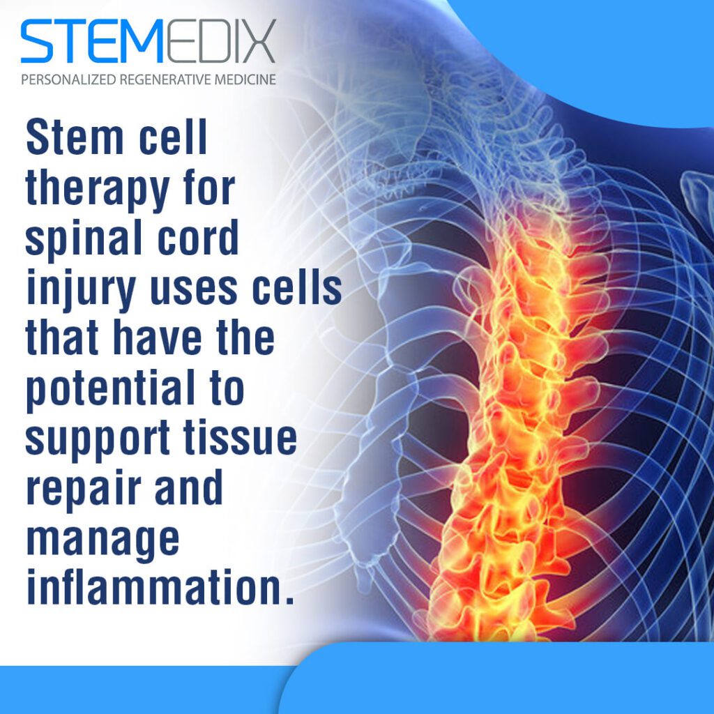


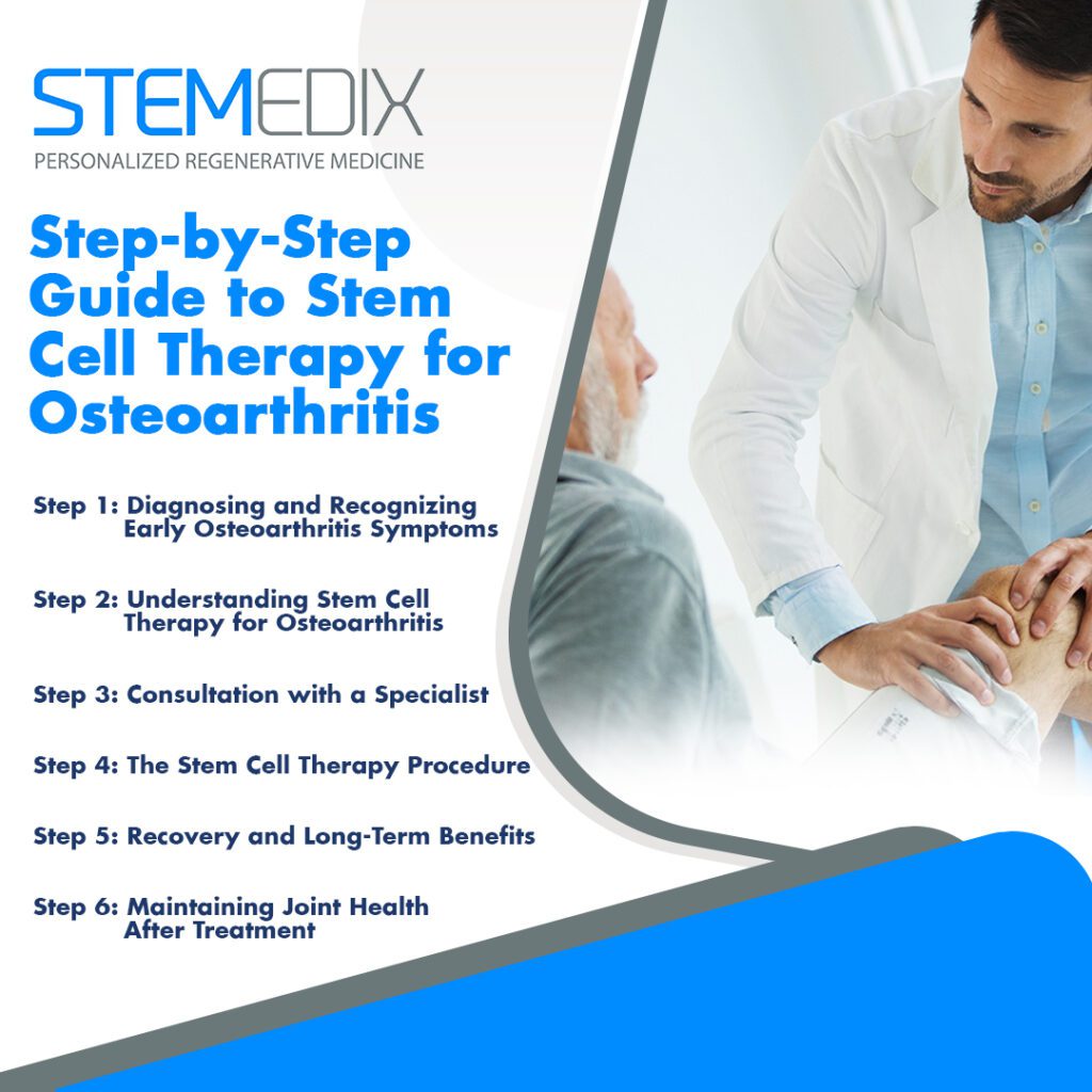
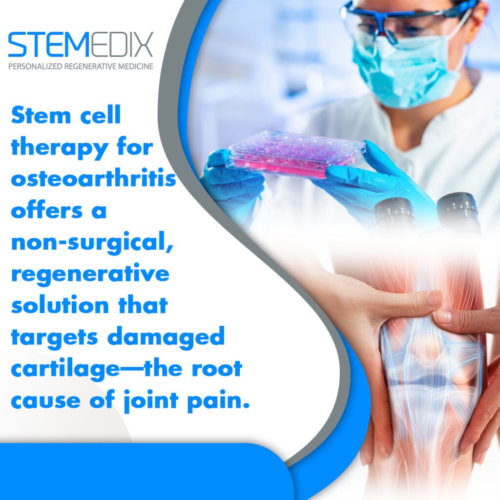


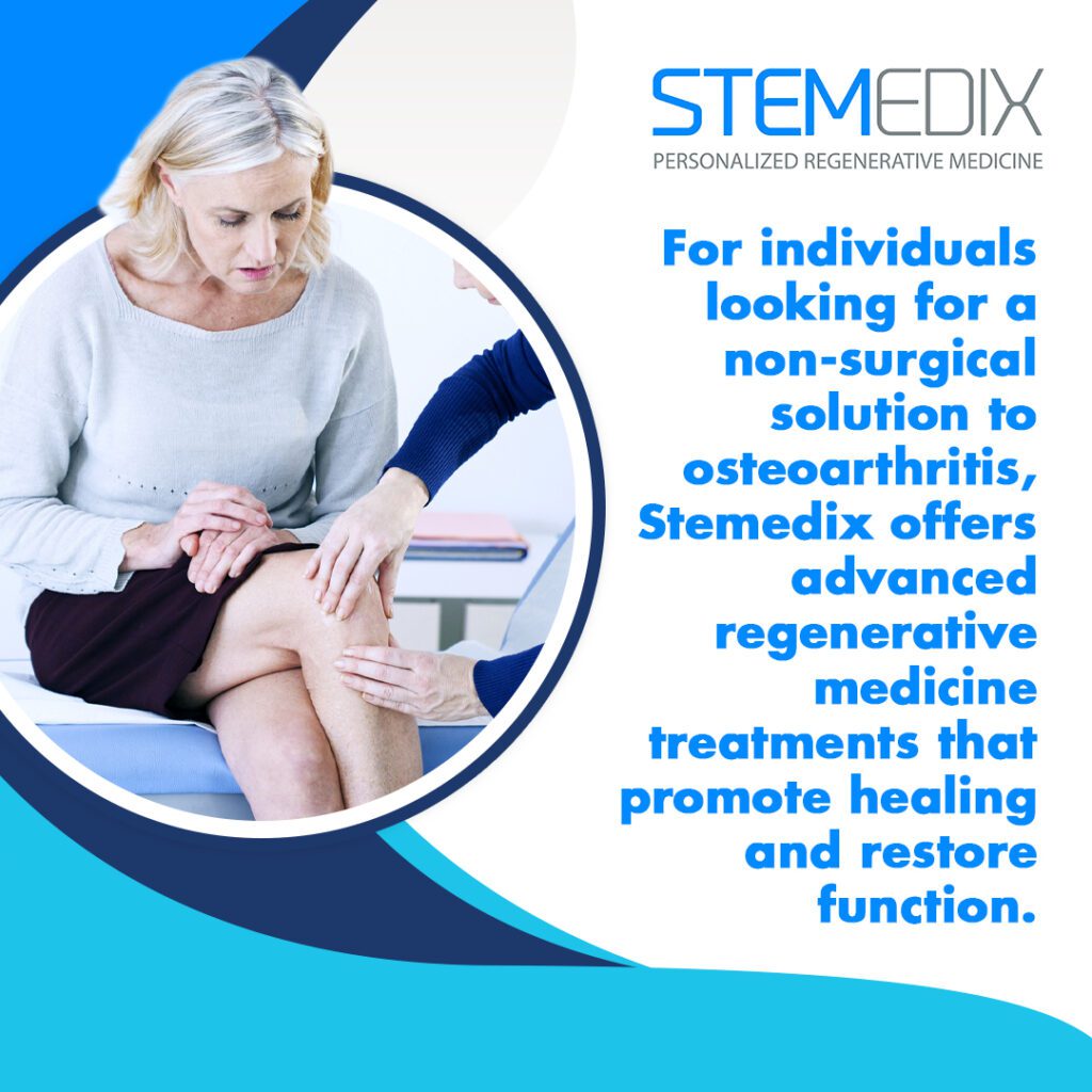

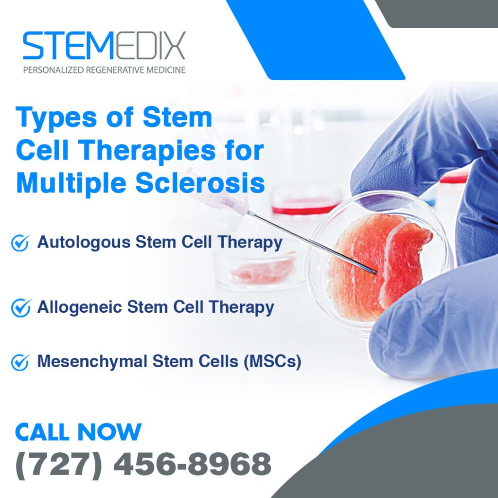

 St. Petersburg, Florida