by admin | Aug 20, 2021 | Peptides
Mitochondria are essential components of each cell in the human body. Responsible for generating the energy required for cells to complete their normal and required functions, large scale or systematic mitochondrial dysfunction, when left untreated often results in low energy, cell damage, and eventually cell death. Mitochondrial disease is most often observed in the heart, liver, muscles, kidney, and brain[1].
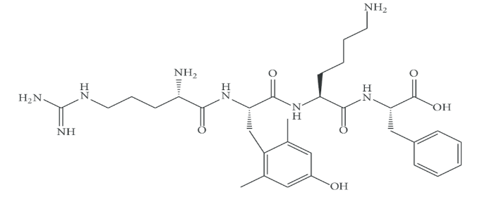
Considering the direct role mitochondria have on neuronal function and considering that perioperative neurocognitive disorder (PND) is one of the most commonly experienced and least understood postoperative complications (especially in elderly patients), Zhao et al. examined the specific role of the mitochondria-targeted antioxidant elamipretide (SS-31) has in preventing mitochondrial dysfunction and synaptic and memory impairment caused by oxidative stress and inflammatory responses.
Considering previous research conducted in this specific area, Zhao et al. hypothesized that elamipretide could offer protection against memory impairment experienced during neuroinflammation specifically by offering protection against mitochondrial dysfunction and by reducing oxidative stress and inflammatory response in the hippocampus.
To test this hypothesis, the authors assigned mice to one of four treatment groups: a control plus placebo group, a control plus elamipretide group, a LPS plus placebo group, or a LPS plus elamipretide group before memory performance and hippocampus-related learning were assessed through a series of open field, Morris water maze (MWM), and fear conditioning tests.
Upon completion of all assays, the authors concluded that Elamipretide:
- Protected the hippocampus against LPS-induced mitochondrial dysfunction by maintaining appropriate levels of mitochondrial membrane potential (MMP) and adenosine triphosphate assay (ATP).
- Reduced oxidative stress and the inflammatory response induced by LPS in the hippocampus (of mice).
- Significantly decreased the death of neural cells within the hippocampus of LPS-treated mice.
- Enhanced the hippocampal brain-derived neurotrophic factor (BDNF) pathway and synaptic structural complexity in mice treated with LPS.
- Prevented the reduction of dendritic spines on hippocampus neurons after LPS treatment.
Although mice treated with LPS demonstrated impaired hippocampus-related learning and memory performance, Zhao et al. concluded that memory impairment caused by LPS can be significantly reduced through the introduction of the mitochondria-targeted antioxidant elamipretide. In addition, elamipretide may also have therapeutic potential when it comes to preventing damage resulting from the oxidative stress and neuroinflammation known to contribute to PND. Considering these findings, the authors call for further exploration into the use of mitochondria as a potential treatment strategy for PND.
Source: (2019, November 20). Elamipretide (SS-31) improves mitochondrial dysfunction, synaptic …. Retrieved from https://jneuroinflammation.biomedcentral.com/articles/10.1186/s12974-019-1627-9
[1] “Mitochondrial Disease Clinic – Clinical Genomics – Mayo Clinic.” https://www.mayoclinic.org/departments-centers/clinical-genomics/overview/specialty-groups/mitochondrial-disease-clinic. Accessed 18 Aug. 2021.

by admin | Aug 13, 2021 | Peptides
While medical knowledge and understanding of the pathophysiology associated with neuropathic pain have increased dramatically over the last 10 years, the medical community is still finding it difficult to control the chronic pain associated with this condition.
Characterized by a wide range of clinical symptoms, including spontaneous pain, allodynia, and hyperalgesia, patients experiencing neuropathic pain and discomfort are also much more likely to experience anxiety, depression, sleep disturbances, and social isolation.
While there are several therapeutic treatment options, including pharmacotherapy, interventional therapies, physiotherapy, and cognitive intervention, less than half of those experiencing neuropathy-associated pain experience adequate pain relief after these treatments. The result is a continuous reduction in quality of life often characterized by severe side effects associated with current pharmacotherapies.
Considering that an important and common feature associated with neuropathic pain is the occurrence of peripheral and/or central inflammation thought to be caused by nerve damage, Dahan et al.’s study examined the role of the innate repair receptor in the treatment of neuropathy.
The innate repair receptor (IRR) consists of the erythropoietin receptor and the β-common (CD131) receptor and is believed to activate anti-inflammatory and tissue repair pathways in response to peripheral nerve injury. Specifically, the IRR appears to be increased in response to injury and as a way to control and/or reduce the typical neurogenic inflammatory response by isolating and destroying the toxins, pathogens, and damaged tissue associated with the injury.
One specific IRR agonist, a small peptide known as ARA 290, appeared to significantly improve symptoms of neuropathy and quality of life in patients with sarcoidosis and with type 2 diabetes (T2D); patients with T2D also showed improved metabolic profile. Treatment with ARA290 for 28 consecutive days demonstrated small nerve fiber regrowth in the cornea, but not in the epidermis.
The authors found that ARA 290 treatment was well tolerated by patients with sarcoidosis and with T2D and produced no significant adverse effects.
Dahan et al. concluded that treatment with IRR offers the potential to reduce tissue damage while also supporting healing and repair occurring as a result of a number of disease processes. Specifically, the peptide ARA 290 has demonstrated the potential to reprogram an area of inflammation and tissue damage into one of healing, growth, and repair.
The authors also point out that while there appears to be potential in the use of IRR for treating neuropathy, clinical observations have been limited to 28 days. As such, the authors also call for longer clinical trials with extended follow-up as a way to assess the full healing potential of IRR activation as disease-modifying therapy.
Source: (n.d.). Targeting the innate repair receptor to treat neuropathy: PAIN Reports. Retrieved from https://journals.lww.com/painrpts/fulltext/2016/08000/targeting_the_innate_repair_receptor_to_treat.2.aspx

by admin | Apr 16, 2021 | Peptides, Neurodegenerative Diseases
Oligodendrocytes are key neural cells responsible for producing myelin sheaths that wrap around neuronal axons in the central nervous system[1]. Considering that thymosin β4’s (Tβ4) ability to promote neurological recovery in a range of neurological diseases has been well established, Chopp & Zhang (2015) propose oligodendrogenesis as the common link by which Tβ4 supports and promotes recovery after neural injury and neurodegenerative disease.
Citing Tβ4’s propensity to alter cellular expression and target multiple molecular pathways involved in neurovascular remodeling and oligodendrogenesis, it warrants further study into Tβ4 as a restorative/regenerative therapy for neurological injury and neurodegenerative diseases.
Traditional treatment of neurological diseases, including stroke, traumatic brain injury (TBI), and multiple sclerosis have typically focused on the reduction of lesions and produced no effective or beneficial long-term therapeutic outcomes. As a result, new proposals suggest renewed focus on therapeutic efforts designed to facilitate the restorative process present after injury and with specific focus on the enhancement of neurovascular recovery resulting in improved neurological recovery.
Among the many benefits of using Tβ4 in the restorative/regenerative therapeutic process is that, unlike neuroprotection treatments that must be introduced to damaged tissue before irreversible damage occurs, this treatment can be administered several days – even weeks – after injury and still stimulate the naturally-occurring regenerative process that has been demonstrated to be beneficial in treating several conditions, including stroke and TBI.
Specifically, Tβ4 promotes the remodeling and restoration of the CNS/PNS post-injury and has been shown to improve neurological recovery by allowing for improved neurovascular plasticity, neurite outgrowth, myelination of axons, as well as increasing the production and release of trophic factors to further support the remodeling of the nervous system.
Multiple animal models have demonstrated that Tβ4 facilitates the restorative neurological process by simulating oligodendrocytes (OLGs) and specifically OLG progenitor cells (OPCs) in the CNS. It appears that Tβ4 expedited multiple pathways of neurological recovery by stimulating tiny non-coding RNAs known as microRNAs to promote the generation, translation, and differential of OPCs and OLGs.
While these findings are promising, what remains yet unknown is specifically how Tβ4 affects, or perhaps more appropriately, influences, microRNAs to communicate specific neurological restorative and regenerative instructions among various cells. The predominant theory emerging from relevant research is that this process of intercellular communication is created and moderated by tiny lipid particles known as exosomes.
Considering the safety of Tβ4 for use in human trials and the potential for Tβ4 to treat neurological injury and degeneration, future clinical trials focusing on Tβ4’s specific influence on exosomes, and as a therapeutic restorative for neurological treatment and regeneration, is thought to hold promising clinical translation for future treatments of neurological disease and injury.
Source: (2015, January 22). Thymosin β4 as a restorative/regenerative therapy for …. Retrieved January 5, 2021, from https://www.tandfonline.com/doi/full/10.1517/14712598.2015.1005596
[1] (2015, October 15). Neuroinflammatory modulators of oligodendrogenesis. Retrieved January 2, 2021, from https://nnjournal.net/article/view/1129

by admin | Mar 26, 2021 | Peptides, Traumatic Brain Injury
With nearly 1.5 million traumatic brain injuries (TBI) occurring each year in the United States and over 69 million more cases of TBI[1] occurring worldwide, TBI continues to be one of the leading annual causes of mortality and morbidity. Carrying with it an annual estimated cost exceeding $56 billion, there has yet to be a viable, effective treatment option for TBI.
As researchers continue to search for an effective pharmacological-based treatment for TBI, recent preclinical studies have found endogenous neurorestoration occurring after TBI. Treatments developed in an effort to improve and/or promote the post-TBI neurorestorative recovery process have also shown to be promising.
Of the potential regenerative peptides identified as part of the TBI recovery efforts, thymosin beta 4 (Tβ4) has been found to exhibit several interesting and promising benefits, including pro-survival and pro-angiogenic properties, protecting tissue against damage, and promoting tissue regeneration.
In addition, previous studies exploring Tβ4 as a potential TBI-treatment option have demonstrated reduced inflammation, improved remyelination, and improved recovery in animal models studying such effects. As a result, the authors of this brief conclude that these observed benefits indicate Tβ4 to hold tremendous potential as a treatment option for TBI.
Since there isn’t one established animal model that can clinically reproduce the significant and multi-layered deficits in cognitive and motor performance experienced as a result of TBI in humans, most TBI studies rely on the controlled cortical impact, or CCI, model to evaluate the efficacy of potential TBI treatments.
Early Tβ4 Treatment May Reduce Cortical lesion Volume and Improve Functional Recovery After TBI
Using the CCI model to evaluate the efficacy of early Tβ4 treatment on spatial learning and sensorimotor functional recovery in rats, the authors demonstrated that rats receiving (Tβ4) treatments 6, 24, and 48-hours after TBI demonstrated significant improvements, especially when compared to rats treated with a vehicle control (in this case, saline). Rats in the Tβ4-treated group also demonstrated reduced hippocampal cell loss and reduced cortical lesion volume at a rate of up to 30%.
As a result of these findings, the authors suggest Tβ4 may encourage neuroprotection even when treatment is provided up to 6-hours after the occurrence of a TBI.
Tβ4 Treatment Provided 24-Hours After TBI Injury May Improve Functional Recovery, But Not Alter Cortical Lesion Volume
The primary intent of any neuroprotective TBI treatment is to reduce the size of established lesion(s). Researchers note that a major, but recurring, limitation of TBI-based treatments is the short timeframe between injury and need for treatment. As such, researchers point out that most preclinical TBI studies found the treatment to be effective only when administered within several hours after experiencing a TBI.
However, when using the CCI model, researchers noted the ability to improve neurological recovery without altering cortical lesion volume; these improvements were observed even when administering Tβ4 treatment more than 24-hours after injury.
Specifically, rats receiving Tβ4 treatment initiated 24 hours post-injury demonstrated significantly reduced cell loss as well as improvement in spatial learning and sensorimotor functional recovery when compared to TBI rats receiving saline treatments.
Tβ4 Treatment Provided 24-Hours After TBI Injury May Promote Neurogenesis
Substantial evidence has repeatedly indicated that all mammals, including humans, have the ability to generate new neurons. Specifically, these neurons originate from neural stem cells in the adult brain and appear to replace neurons that die regularly and in specific areas of the brain.
Considering this information, scientists introduced bromodeoxyuridine (BrdU), a thymidine analogue, into the DNA of dividing cells with the goal of improving neurogenesis. When compared to sham controls, this process demonstrated a significant increase in the presence of BrdU-positive cells 35 days after TBI.
Additionally, Tβ4 treatment appeared to further increase the number of BrdU-positive cells when compared to saline controls. These findings were further supported by the authors’ data confirming neurogenesis increases in TBI rats receiving Tβ4 treatment. The authors suggest that these findings indicate Tβ4 treatment promotes new cells to differentiate into brain-specific neurons.
Tβ4 Treatment Provided 24-Hours After TBI Injury May Promote Angiogenesis
Research demonstrates normal adult brains contain a stable vascular system; however, the vascular system becomes active in response to several pathological conditions, one of them being TBI. Findings indicate that Tβ4 treatment applied to specific areas of the brain associated with TBI, including the cortex, demonstrate significant increases in the observed vascular density of these specific areas, especially when compared to a control group receiving a saline treatment.
The presence of increased vascular density in these areas of the brain is thought to be closely related to improved recovery from TBI and specifically enhanced neurogenesis and synaptogenesis. As a result, the authors of this brief suggest further study to attain a better understanding of neurovascular molecular mechanisms with a specific focus on developing angiogenic and neurogenic therapies for TBIs.
Considering the above findings pertaining to the potential benefits of Tβ4 treatment for TBI and the fact that administration of Tβ4 has proven safe and well-tolerated in both animals and humans, the authors call for further investigation of molecular mechanisms that contribute to enhanced Tβ4-mediated neuroprotection and neurorestoration.
Source: (n.d.). Neuroprotective and neurorestorative effects of Thymosin beta 4 …. Retrieved February 4, 2021, from https://www.ncbi.nlm.nih.gov/pmc/articles/PMC3547647/
[1] “Estimating the global incidence of traumatic brain injury – PubMed.” 1 Apr. 2018, https://pubmed.ncbi.nlm.nih.gov/29701556/.

by admin | Jan 13, 2021 | Health Awareness, Peptides
What Is BPC-157 Peptide & What Does It Help?
BPC stands for “body protective compound.” BPC-157, in particular, is a synthetic peptide with 15 amino acids. It has been derived from digestive proteins and is largely used to prevent stomach ulcers. Recently, however, the supplement has been shown to offer many other benefits.
For instance, BPC-157 has been found to help stabilize the microbiome or healthy balance of bacteria in the gut. It also controls blood pressure function by interacting with the nitric oxide pathway. In addition, it promotes growth factors, unlocking the regenerative potential to help the body heal and empower its systemic repair response.
Across various bodies of research, BPC-157 has been linked to a wide range of healing benefits. In addition to repairing blood flow, it has been shown to:
- Help with burns
- Increase collagen production
- Aid in the healing of sprains, tears, and other muscle injuries
- Help with inflammatory conditions, such as arthritis
- Aid in weight loss
- Repair ligament and tendon-to-bone injuries or damage
- Protect the cardiovascular system
- Reduce damage from drugs and the effects of corticosteroid injections
- Improve responses to allergens and viruses
- Boost brain health and mood
- Protect scar tissue formation
BPC-157 is often taken via oral capsule, especially when its goal is to treat stomach or intestinal issues. It can also be injected subcutaneously or intramuscularly and used as a nasal spray. The best delivery method may vary based on the patient and their unique concerns, so be sure to discuss your goals with a medical professional when deciding to introduce BPC-157 into your supplement regimen.
For more information to discover if this peptide may be a benefit for you, please call our team at 800-531-0831.

by admin | Jul 6, 2020 | Peptides
Multiple sclerosis is a challenging disease for patients, caregivers, and physicians. MS takes function away from patients, causing them to lose the ability to walk, to feel, or to see. The symptoms of MS vary from patient to patient. In fact, the symptoms of multiple sclerosis can even change in the same patient over time. Wherever MS destroys covering on nerve cells, there are the symptoms of the disease. These functional deficits are challenging for caregivers who provide support for patients. MS is also challenging for physicians because it is a difficult disease to treat.
Several disease-modifying treatments have been released over the past decade that has changed the course of MS for some patients. None is a cure. As such, researchers are continually looking for new treatments for multiple sclerosis.
One of the more promising potential MS treatments is thymosin beta 4. Thymosin beta 4 is a small protein (i.e. peptide) that the body produces naturally. When the protein is injected into animals, it produces regenerative and restorative benefits such as improved wound repair and blood vessel and nerve regeneration.
When scientists want to test new treatments for MS, they often use a model of the disease called experimental autoimmune encephalomyelitis (EAE). When researchers cause EAE in mice, it causes the mice to experience symptoms and changes that are quite similar to MS in humans. Once mice are given EAE, scientists can then see if a particular treatment can make the mice better.
This is precisely what Dr. Zhang and co-researchers accomplished; they caused experimental MS in mice and treated them with either thymosin beta 4 or placebo (saline) injections. Mice began recovering from EAE as early as 11 days after treatment with thymosin beta 4. Thymosin beta 4 treatment reduced the severity of experimental MS in mice by about half compared to those who received a placebo. The statistically significant benefit lasted at least 30 days after thymosin beta 4 treatment.
The researchers performed additional experiments to determine how thymosin beta 4 improved function in mice with EAE. Based on experiments in mouse nerve cells, thymosin beta 4 appears to reduce inflammation and increase oligodendrogenesis, which is the growth of new cells the replace the covering on nerves.
While this work was performed on mice and will need to be confirmed in humans, it is an exciting lead in the development of new therapies for MS. Since thymosin beta 4 appears to be safe, this small protein may one day be among the disease-modifying treatments for MS.
Reference: Zhang, J., et al. (2009). Neurological Functional Recovery After Thymosin Beta4 Treatment in Mice with Experimental Auto Encephalomyelitis. Neuroscience. 2009 Dec 29; 164(4): 1887-1893.


 St. Petersburg, Florida
