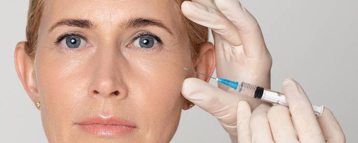
by admin | Sep 2, 2019 | Aesthetics, Stem Cell Therapy
No matter how young we may feel, our aging skin often gives our age away. As we age, we lose important proteins in the skin like collagen and elastin. We also lose a thin layer of fat that lies just below the skin. Without this fat and protein, the skin sags and loses its elasticity (“bounce back”) appearing thin and wrinkly.
One of the main skin rejuvenation techniques is to inject fat into aging skin. In its most basic form, a bit of fat is removed from the patient via liposuction and injected into an area of aging skin. These transplanted fat cells give the skin a plumper appearance and seem to turn back the clock on skin aging. However, mature fat cells do not last forever, and many people need to have repeated skin rejuvenation procedures to maintain the effect.
More recently, however, physicians have realized that they achieve better results when they include stem cells in their fat grafts. The mature fat cells provide a beauty benefit immediately, while stem cells help maintain the effect longer. Some physicians have taken the procedure so far as to only inject stem cells and remove mature fat cells completely. While this approach seems reasonable, is unclear whether the stem cell-only approach is actually superior.
While the benefit of fat grafts that include stem cells is widely known, it is unclear how the process works. By understanding the process, researchers hope to learn what provides the strongest antiaging effect. To help them better understand the skin rejuvenation process of fat-derived stem cells, researchers performed liposuction on several patients and then reinjected fat cells and stem cells into areas of aging skin. Three months later, the researchers took samples of the skin to see what effect the combination of fat cells and stem cells had on the skin structure.
While the specific results are quite complex, researchers found that a fat graft that included adipose-derived stem cells improved the elasticity of the skin by creating what is known as an “oxytalan elastic network.” These fibers helped to smooth out the skin and allowed it to retain more moisture than it did before skin rejuvenation treatment.
Perhaps most importantly for patients, the skin rejuvenation procedure left patients with fuller, younger-looking skin.
The researchers concluded that the most beneficial skin rejuvenation approach is to inject a combination of fat cells and stem cells derived from fatty tissue. The benefits were the same as those obtained by injecting stem cells alone. The researchers argue that the combination approach provides a good clinical benefit with the added benefits of being easier to perform, cheaper, and less prone to complications.
Reference: Charles-de-Sá, L. et al. (2015). Antiaging treatment of the facial skin by fat graft and adipose-derived stem cells. Plastic and Reconstructive Surgery. 2015 Apr;135(4):999-1009.
by admin | Aug 29, 2019 | Age Management, Mesenchymal Stem Cells, Stem Cell Therapy
Cognitive aging describes the changes to our ability to think, remember, and process information that occurs as we age. Cognitive aging begins in adulthood and progresses—if not accelerates—in old age. Over time, the speed at which we process information in the brain slows down, our ability to pay and maintain attention decreases, and we have a harder time making and recalling new memories. While some view cognitive aging as normal because it occurs in all of us, others acknowledge that cognitive aging is something that interferes with a person’s ability to function and diminishes the quality of life.
Currently, there are very few things that can slow cognitive aging and essentially nothing that can reverse it. Physical exercise, mental activity, and a healthful diet can modestly preserve cognitive function as we age. However, once aging occurs in the brain, there is nothing that we can do—currently—to change it.
Some innovative scientists are trying to change that, however. They are focusing on the changes in the brain that take place during aging and using stem cells to reverse that process.
A group of neuroscientists focused their efforts on memory and on the hippocampus, which is the main region of the brain that is responsible for memory. Researchers collected clinical-grade, mesenchymal stem cells taken from human umbilical cords and infused them into aging mice. Aging mice received stem cell treatment once every two weeks for several months.
After three months of treatment with umbilical cord-derived mesenchymal stem cells, mice showed significant improvement in learning and memory tests. Treated mice also had a remarkably improved function in the hippocampus. Surprisingly, stem cell treatment also created new brain cells (i.e. neurogenesis). Indeed, stem cell transplantation in aging mice actually reversed changes in the brain associated with cognitive aging.
These results were conducted in mice and not in humans, however, this research offers a strong foundation for conducting clinical human studies. If these improvements in memory and brain health could be shown in humans, it would be a groundbreaking study. Even in its current form, this research is an exciting breakthrough for the fields of stem cell medicine, neuroscience, and the neurobiology of cognitive aging. It suggests that mesenchymal stem cells may one day be able to reverse cognitive aging.
Reference: Cao N. et al. (2017). Clinical-grade human umbilical cord-derived mesenchymal stem cells reverse cognitive aging via improving synaptic plasticity and endogenous neurogenesis. 2017 Aug 10;8(8):e2996.
by admin | Aug 22, 2019 | Erectile Dysfunction, Stem Cell Therapy
The little blue pill brought erectile dysfunction from out of the shadows and into our shared awareness. Erectile dysfunction affects millions of men and, in turn, their sexual partners. The condition can undermine a man’s sense of self-worth, self-esteem, and masculinity. While the little blue pill has been instrumental in getting men with erectile dysfunction to ask their doctors about treatment, that same pill does not work for every man. Indeed, countless men fail to achieve successful erections even after taking oral erectile dysfunction medication.
In one of the first clinical studies of its kind, urologists at a medical practice in Florida tested the effects of mesenchymal stem cell treatments in men with erectile dysfunction. They selected eight men with erectile dysfunction who could not achieve erections even after oral medications. The men received mesenchymal stem cells that were derived from human placenta (also known as afterbirth). The urologists then followed the men for six months after treatment, testing blood flow, penile size, and erectile function.
The men treated with mesenchymal stem cells had a statistically significant increase in penile blood flow at six weeks, three months, and six months after treatment. Three men were able to achieve erections within three months of treatment without oral erectile dysfunction medication. After stem cell treatment, four other men were able to achieve erections with low-dose oral erectile dysfunction medication (which had previously been ineffective).
Importantly, the treatment was well-tolerated by all men in the study, which is an important milestone for continuing this research.
The study is potentially groundbreaking as it opens the door to larger clinical studies in men with erectile dysfunction. Indeed, nearly two dozen clinical trials are now studying the effects of stem cell treatment for erectile dysfunction. These early results are exciting, and offer hope to men with erectile dysfunction, especially those for whom oral medications have failed.
Reference: Levy, JA (2016). Determining the Feasibility of Managing Erectile Dysfunction in Humans With Placental-Derived Stem Cells. The Journal of the American Osteopathic Association. 2016 Jan;116(1):e1-5.
by admin | Aug 19, 2019 | Exosomes, Stem Cell Research, Stem Cell Therapy
Given the limitations of several conventional methods to treat a wide variety of diseases and injuries, stem cell therapy has begun to gain in popularity. The evidence supporting the field of Regenerative Medicine, which involves using stem cells to regenerate healthy, functional tissue, has indeed been accumulating in recent years.
There are a number of different types of stem cells that have been explored for their therapeutic potential. Mesenchymal stem cells have become a preferred option for therapy because of their ability to differentiate into several different types of adult tissue and to be transplanted safely and effectively into patients.
One-way mesenchymal stem cells confer their therapeutic benefits is through paracrine effects that are achieved by the secretion of extracellular vesicles, some of which are exosomes. Exosomes are between 30 and 100 nanometers (nms) in diameter and exist in blood, cerebrospinal fluid, and other bodily fluids.
A recent review, published in Cell Transplantation, covered research showing that mesenchymal stem cell exosomes are therapeutically advantageous for the management of several conditions, including Parkinson’s disease, osteoarthritis, and stroke.
The review discusses, for instance how in models of Parkinson’s disease, exosomes have been shown to provide neuroprotection. MSC-derived exosomes also appear to inhibit inflammation in the context of osteoarthritis and also to stimulate repair in damaged tissue. Further, specific exosome biomarkers, miR-9 and miR-124, have proven to be promising in diagnosing the severity of stroke.
Based on recent research covered in this review, stem cell-derived exosomes have significant therapeutic potential. Though this review focuses specifically on the relevance of exosomes in Parkinson’s disease, osteoarthritis, and stroke, exosomes will likely provide benefits for patients in a variety of contexts and will prove to be an important part of Regenerative Medicine.
Reference
Chang, Y-H, et al. (2018). Exosomes and stem cells in degenerative disease diagnosis and therapy. Cell Transplantation, 27(3), 349-363.
by admin | Jul 12, 2019 | Age Management, Exosomes, Stem Cell Therapy, Umbilical Stem Cell
As we age, the appearance and structure of our skin changes. This is plain for all to see—we can usually estimate a person’s age simply by looking at their skin. Young skin is full of molecules that keep it thick, plump, and supple, such as collagen and elastin. Over time, the skin produces less and less of the substances. Consequently, aging skin is thinner and it loses its strength and elasticity. As such, the skin develops fine lines and deep wrinkles. It also becomes lax and begins to sag.
Scientists know that the quantity of collagen, elastin, and other proteins make the difference between young and old skin. Not surprisingly, doctors have been trying for decades to increase the levels of these molecules in the skin in an effort to reverse the signs of aging skin. Some approaches work for short periods of time. For example, laser and intense pulsed light treatments can stimulate the skin to produce these youthful molecules. Another approach is to directly inject collagen and other substances into the skin. The Holy Grail of skin rejuvenation, however, is to find a way to make the skin naturally produce more of these substances. Recent research suggests that stem cells could be the answer.
Researchers collected mesenchymal stem cells from umbilical cords. This tissue is removed and discarded after a woman gives birth to a baby. The scientists then collect the tiny sacs called exosomes from these umbilical cord stem cells. Exosomes are densely packed with proteins, RNA, and other important molecules that are important for growth in rejuvenation. The researchers then simply applied these exosomes to samples of human skin to see if they could influence skin rejuvenation.
The first remarkable finding of this research was that exosomes taken from umbilical cord mesenchymal stem cells were absorbed into human skin. Why is this important? Because it means that if exosomes are used as a potential treatment, they can be placed on the skin rather than injected into the skin. The second remarkable finding is that exosomes, once they cross into the skin, are taken up by skin cells (human dermal fibroblasts). Once inside the skin cells, the exosomes take over the cells, in a way. They prompt the cells to produce more collagen and elastin than normal. The exosome-treated skin cells also attract other cells to the skin. We know from other work that more collagen, more elastin, and more cells within the skin leads to plumper, fuller, more elastic skin.
While clinical trials are needed to confirm this research, this work strongly suggests that the exosomes from umbilical cord-derived mesenchymal stem cells have the ability to rejuvenate human skin. Perhaps most impressively, these potential skin rejuvenating exosomes can be applied topically, such as within a cream or ointment. Thus, patients could receive the potential benefits of this treatment, while avoiding painful injections. Again, more work needs to be done before this research becomes a routine treatment, but the results are quite promising.
Reference: Kim, YJ. et al. (2017). Exosomes derived from human umbilical cord blood mesenchymal stem cells stimulate rejuvenation of human skin. Biochemical and Biophysical Research Communications. 2017 Nov 18;493(2):1102-1108.
by admin | Jul 8, 2019 | Exosomes, Osteoarthritis, Stem Cell Research, Stem Cell Therapy
Osteoarthritis is the most common form of arthritis. About one in 10 people will develop osteoarthritis at some point in their lifetimes. As the condition progresses, synovial membranes and cartilage break down. Osteoarthritis causes people to experience joint pain, joint stiffness, and restricted movement. Knees, hips, and hands are common sites for arthritis, though people can experience the condition in virtually every joint in the body including joints and spine. Mild osteoarthritis may be nothing more than an annoyance, but moderate and severe osteoarthritis can diminish a person’s quality of life and cause substantial suffering and disability.
Despite the commonness of osteoarthritis, there are very few effective treatment options. People may take pain medications to help cope with discomfort; however, taking these medications every day can lead to unwanted side effects. Physical therapy, braces, walking aids, and exercise may have some effect, but their benefit is unpredictable, i.e., these approaches work for some people and not others. The only definitive treatment for osteoarthritis is to replace the joint with an artificial one; however, orthopedic surgery is expensive, associated with a long recovery, and is usually only an option after patients have suffered pain and disability for a long period of time.
Ideally, osteoarthritis treatment would be focused on restoring the structure of the damaged joint itself. For a time, physicians were hopeful that glucosamine and chondroitin could do this. These two substances are diminished in osteoarthritis, so the concept was to replace them and hopefully rebuild damaged joints. While initial clinical studies seemed to suggest glucosamine and chondroitin were helpful, larger, high-quality clinical trials failed to show any benefit. Nevertheless, the goal of repairing joints damaged by osteoarthritis is still a top goal. Now, however, scientists have turned to stem cells instead of small molecules.
Stem cell researchers are particularly interested in the small sacs released by stem cells called exosomes. Exosomes contain proteins, RNA, cytokines, and other substances that are important for tissue regeneration. Exosomes are apparently responsible for most of the clinical benefit of stem cells in the first place. Moreover, it is much more convenient for doctors to use exosomes as treatment rather than stem cells themselves.
To this end, researchers have been studying the effects of stem cell exosomes. One research group recently published a report studying the effects of exosomes that were collected from synovial membrane stem cells and compared them to exosomes taken from induced pluripotent stem cells. Synovial membrane stem cells are an obvious choice for treating osteoarthritis since synovial membranes are one of the substances that break down in the disease. However, synovial membrane stem cells are difficult to obtain. It is far more practical to use induced pluripotent stem cells since there is a virtually limitless supply of them.
The researchers compared exosomes taken from these two types of stem cells in mice with experimental osteoarthritis. Not only did the scientists show that exosomes taken from pluripotent stem cells were highly effective in treating osteoarthritis, these exosomes were even more effective in treating osteoarthritis then exosomes taken from synovial membrane stem cells. Exosomes from pluripotent stem cells not only repaired synovial membranes but they also helped replace cartilage by stimulating chondrocytes (cartilage cells).
Of course, this work will need to be confirmed in humans with osteoarthritis; however, the results of this animal research are very encouraging. In short, this research suggests that exosomes taken from induced pluripotent mesenchymal stem cells may be a convenient and effective way to treat osteoarthritis in the future.
Reference: Zhu, Y. et al. (2017). Comparison of exosomes secreted by induced pluripotent stem cell-derived mesenchymal stem cells and synovial membrane-derived mesenchymal stem cells for the treatment of osteoarthritis. Stem Cell Research and Therapy. 2017 8:64.


 St. Petersburg, Florida