by admin | Nov 14, 2018 | Stem Cell Research, Stem Cell Therapy
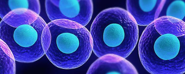
Evidence has been accumulating for years showing how stem cells can serve therapeutic functions. Much of this research focuses on how stem cells can be applied to damaged tissue to help regenerate the area. Because stem cells can differentiate into a wide variety of cell types, they can be widely utilized to repair distinct types of tissue. However, a recent paper published in the World Journal of Stem Cells has described how stem cells can also be used to carry therapeutic agents to tissues and organs to help with regeneration.
Stem cells are good candidates for delivering genes, proteins, and small molecules to areas of interest because they have an innate ability to migrate to sites of injury. One challenge for using stem cells for this type of therapeutic delivery is how to load the stem cells with the therapeutic agents. There are pros and cons for the techniques that have been investigated.
Polymeric nanoparticles, are FDA approved and are versatile, uploaded efficiently, and biocompatible. However, it is hard to control the release of the therapeutic agent from the stem cells. Magnetic nanoparticles are not associated with high levels of toxicity and are efficient with loading. However, they can induce oxidative stress in carrier cells.
Silica nanoparticles have quick uptake, are non-toxic, stay within cells for a long time, and are versatile. However, their tendency to stay within cells for a long time can sometimes be a disadvantage when the agent needs to be cleared.
Liposomal nanoparticles are relatively easy to manufacture and are versatile in their therapeutic agent delivery. However, these nanoparticles are less efficient at uptake and need higher concentrations of the therapeutic agent loaded, which can be toxic to cells.
Once stem cells are loaded with bioactive molecules, there are a few ways that they can be guided toward target organs. For instance, they can be systemically infused so that they can migrate to their target areas trough blood flow.
Further research will help to clarify how well stem cells can be used to help deliver therapeutic agents to damaged or impaired tissue. Investigation into the different nanoparticles, stem cells, and potential therapeutic applications will help us better understand the extent to which stem cells can be used in regenerative medicine.
by admin | Oct 30, 2018 | Studies, Stem Cell Research, Stem Cell Therapy
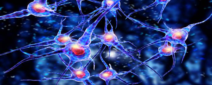
Spinal cord injury is the second leading cause of paralysis in the United States. When the spinal cord is severely injured, nerve cells in the spinal cord are damaged or destroyed. Also, a sort of scar forms in the affected area, which prevents nerve signals from traveling between the brain and the extremities. Consequently, people who sustain spinal cord injuries suffer from paralysis. The degree of paralysis depends on the location of the spinal cord injury; injuries higher on the spinal cord such as the neck or upper back area can lead to paralysis of all four limbs, for example. In almost all cases, the paralysis is permanent once it occurs, because nerve cells in the spinal cord do not regenerate.
Because spinal cord injuries are common and the consequences are usually permanent, researchers have been aggressively and tirelessly researching ways to treat this condition. One approach is to try to form new nerve cells in the spinal cord using stem cells. Mesenchymal stem cells can become new nerve cells given the right set of circumstances. Unfortunately, simply injecting mesenchymal stem cells into patients with severe spinal cord injuries cannot reverse paralysis. On the other hand, using exosomes from mesenchymal stem cells may be the push that stem cells need to become nerve cells in the spinal cord.
Exosomes are tiny packets of cellular material released by stem cells. They contain a variety of potentially beneficial substances; perhaps the most important in cell regeneration is micro RNA (miRNA). miRNA can cause complex changes in cells that simple drugs, proteins, or even regular RNA cannot. Researchers cannot easily deliver miRNA to where it is needed in the body, but exosomes taken from stem cells can deliver miRNA right where it needs to be.
Researchers collected human mesenchymal stem cells and placed them in an environment that would cause them to become nerve cells. But instead of simply using the stem cells directly, they instead collected the exosomes from those stem cells. Those exosomes could then be used to prompt mesenchymal stem cells to become nerve cells. Simply put, the exosomes drove the process more efficiently than the stem cells alone.
What does this all mean? Exosomes taken from the mesenchymal stem cells could eventually be used to treat spinal cord injury. Those special exosomes would magnify the nerve cell-creating effect, perhaps restoring nerve cell function to a damaged spinal cord. Considerable research needs to be done before this possibility becomes a clinical reality, but this knowledge helps researchers design targeted experiments in the future.
by admin | Oct 18, 2018 | Stem Cell Research, Stem Cell Therapy, Studies
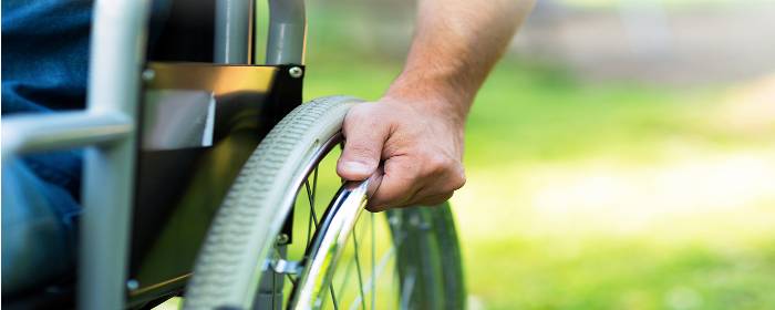
A couple of weeks ago, scientists published findings showing that implanting human stem cells that are embedded within the engineered tissue can lead to the recovery of sensory perception in rats. The recovery of sensory perception is also accompanied by healing within the spinal cord and the ability to walk independently. The stem cells used in this experiment were collected from the membrane lining the mouth.
These results help demonstrate the potential for stem cells to help with spinal cord injuries but also point to the utility of combining stem cells with other factors to enhance their therapeutic effects. In this case, the researchers used a 3-dimensional scaffold to enable stem cells to attach and to stabilize them in the spinal cord. By adding growth factors, such as human thrombin and fibrinogen to the engineered tissue scaffolding, the researchers also increased the chances that attached stem cells would grow and differentiate.
The researchers compared the effects of their stem cell implants in paraplegic rats with the effects of adding no stem cells. Whereas the control rats who did not receive stem cells did not experience any improvement in mobility or sensation, 42% of the rats that did receive stem cells became better at supporting their weight on their hind limbs and at walking.
While these results are pre-clinical and do not apply directly to humans, the researchers conclude that further research is warranted. Given the positive impact of stem cells on the spinal cord in animals, it is reasonable to assume that stem cells may also benefit the human spinal cord. Further research will help clarify whether these stem cells can be adequately used to help treat patients with paraplegia.
by admin | Oct 11, 2018 | Stem Cell Research
Myocardial infarction, also known as heart attack, can be a devastating or even deadly event. It occurs when blood flow in one or more coronary arteries is blocked. Since coronary arteries supply blood to the heart muscle, a blockage in a coronary artery prevents oxygen and nutrients from reaching heart tissue.
While the heart can sustain short periods of time without oxygen or nutrients, heart cells become dysfunctional and die if blood flow is not restored within several hours. While clot-busting drugs, percutaneous intervention (PCI), and balloon angioplasty have provided a way to restore blood flow to the heart during a heart attack, once heart cells die there is no way to bring them back. Since most heart tissue is cardiac muscle, dead heart tissue cannot participate in the contraction or squeezing of the heart during a heartbeat. Thus, people who have survived a heart attack are often left with poor heart function (e.g. congestive heart failure).
Stem cell researchers have begun to question whether heart tissue destroyed during a heart attack is necessarily gone forever. Research is beginning to show that stem cells given after myocardial infarction are able to improve the squeezing power of the heart. By extension, stem cell treatment is able to improve the abilities heart to pump blood throughout the body.
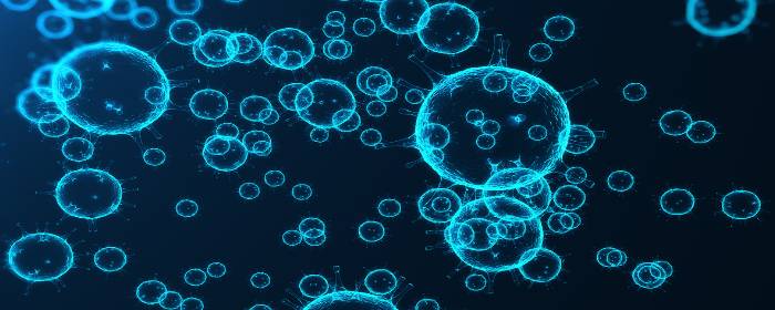
Researchers initially assumed that it was the stem cells themselves that became new heart cells, replacing dead and dysfunctional heart tissue. While there is evidence that this occurs, it seems that stem cells play an even bigger role in heart tissue repair than simply becoming new heart cells. Stem cells release small packets of a material called exosomes and microvesicles. Exosomes and microvesicles hold proteins, cytokines, chemokines, growth factors, DNA, messenger RNA, and micro RNA. Researchers now believe that these materials hold the true power of stem cells in cardiac repair and regeneration.
Various types of stem cells produce exosomes that could potentially help repair a damaged heart. While cardiac stem cells may seem like an obvious source for these exosomes, induced pluripotent stem cells and mesenchymal stem cells are also capable of releasing exosomes that are potentially beneficial in cardiac repair.
Stem cells—or more accurately the exosomes contained within the stem cells—help repair damaged heart tissue in several ways. Stem cell-derived exosomes contain factors that promote the survival of vulnerable heart cells and cells that are dysfunctional after a heart attack (but not dead). Exosomes also help new blood vessels to form in and around the damaged heart muscle in a process called angiogenesis. These new blood vessels deliver oxygen, nutrients, and molecules that help support the growth and function of heart tissue. Exosomes also appear to promote a healthy immune system response after a heart attack, rather than a destructive inflammatory reaction. In other words, the materials found in exosomes guide the immune system to clear away damaged tissue without creating extensive fibrotic (i.e., tough, nonfunctional) tissue.
While most clinical trials thus far have studied the effects of stem cells directly infused into humans after myocardial infarction, exosomes are rapidly becoming the focus of future clinical trials in this area.
by admin | Sep 24, 2018 | Stem Cell Research, Stem Cell Therapy
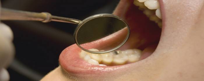
The application of stem cells to treat health disorders, diseases, and injuries has been rapidly expanding in recent years. The breadth of their application comes from the fact that stem cells are undifferentiated and can, therefore, differentiate into all sorts of cells with different specialized functions and therefore have an enormous number of potential ways that they can improve health. A review published in Frontiers in Physiology covers the way stem cells can be used for therapy of oral diseases.
According to the authors of the article, adult stem cells and induced pluripotent stem cells are the best types of stem cells to use to treat oral and maxillofacial defects. There are pros and cons associated with adult stem cells, including both autologous and allogeneic stem cells, as well as with induced pluripotent stem cells. For instance, whereas autologous stem cells can modulate the immune system, allogeneic stem cells appear helpful for malignant diseases, and induced pluripotent stem cells are unlimited in terms of their source and do not involve any ethical issues.
There are a number of potential sources for treating oral disease, including tooth germ progenitor cells, dental follicle stem cells, salivary gland stem cells, stem cells of the apical papilla, dental pulp stem cells, inflamed periapical progenitor cells, among others. While adults stem cells can differentiate directly into specialized cells or can be turned into induced pluripotent stem cells, induced pluripotent stem cells can be driven to differentiate into specialized cells.
Clinical trials have been undertaken to study the ways in which stem cells can address a number of oral diseases, including bone diseases, dental pulp diseases, eye diseases, facial diseases, and periodontal diseases, as well as tooth extraction. The strategies for treating oral disease with stem cells involve sorting and expanding the stem cells outside of the body, mixing them with materials and factors that help them grow, and implanting them into the impaired region.
Future research will help to delineate the different ways in which certain types of stem cells can best be used to address individual oral diseases. Studies will also help to uncover the specific types of stem cells that are best for specific diseases and the protocols that should be used to reap the greatest benefits for patients.
by admin | Sep 13, 2018 | Stem Cell Research, Stem Cell Therapy, Studies
Patients usually recover from bone fractures with the right treatment, but sometimes the bone fails to heal because new tissue does not form and connect the broken pieces properly. Delayed union refers to cases where the bone takes longer than usual to heal, and nonunion refers to cases where the bone does not heal. In approximately 5 to 10 percent of cases of a fractured bone, delayed union or nonunion occurs. These conditions are associated with long-term pain and discomfort, and though can be addressed through surgical treatments, these interventions do not always lead to long-term healing.
In recent years, researchers have begun exploring the potential for mesenchymal stem cells to help address these important challenges of delayed union and nonunion. A review of the potential for these stem cells to help in these cases where fractures do not properly heal was recently published in the Journal of Biomedical Materials Research.
Mesenchymal stem cells are helpful in bone healing because they differentiate well and can differentiate into different cell lineages that are all important for bone formation, growth, and maintenance. These cell types include chondrocytes, osteoblasts, myoblasts, and adipocytes.
According to the authors of the review, mesenchymal stem cells can be used in conjunction with extracellular matrix scaffolds and biological adjuvants that promote growth, differentiation, and blood vessel formation, to help in the bone healing process when the delayed union or nonunion occurs. Future research will help to determine the best ways that mesenchymal stem cells can be used in combination with bioengineering strategies to help patients whose bone fractures do not heal or do not heal properly.

 St. Petersburg, Florida