by admin | Jan 11, 2017 | Studies
A group of scientists, led by Jin Nam at the University of California-Riverside, have found a new way to optimize the use of stem cells for orthopedic purposes. The technique involves strategically using biomechanical forces to create the specific cell type needed to repair orthopedic tissues.
The normal development of bone and cartilage critically depend on mechanical stimulation, yet the ideal level of mechanical stimulation differs for cells that make up bone and cells that make up cartilage. The researchers therefore reasoned that employing the right level of mechanical stimulation could help stem cells differentiate into the cell type needed for specific individual injuries.
A certain type of stem cell, called the mesenchymal stem cell (MSC) has been shown to be capable of regenerating musculoskeletal tissues. In addition, when MSCs obtained from the patient are used to regenerate that patient’s musculoskeletal tissues, tissue rejection tends to be prevented because the immune system recognizes the transplanted cells as the body’s own cells. Nonetheless, stimulating the stem cells to form tissue has relied mainly on biochemical and biophysical phenomena, which can present a variety of challenges. To avoid those challenges, scientists hoped that they could use biomechanical stimulation for the same purpose.
In their experiment, Nam and his colleagues showed that different levels of biomechanical force could indeed lead MSCs to differentiate into osteoblasts and chondrocytes, the cells that make up bone and cartilage, respectively. These promising results suggest that combining these types of stem cells with targeted biomechanical stimuli will enable better repair of a variety of types of orthopedic damage.
Going forward, researchers hope to better understand the details of how biomechanical cues can determine the fate of stem cells and to use this information to induce stem cells to differentiate into the desired cell type, according to the particular injury. This experiment conducted at the University of California-Riverside helps shed more light on the continued value of stem cells in medicine, as well as the potential benefits of combining these cells with approaches that can help personalize the treatments offered.
Stem cells could revolutionize therapeutic strategies for cartilage repair. Find out more.
Reference
Horner, C.B. et al. (2016). Magnitude-dependent and inversely-related osteogenic/chondrogenic differentiation of human mesenchymal stem cells under dynamic compressive strain. Journal of Tissue Engineering & Regenerative Medicine. doi: 10.1002/term.2332
by admin | Jan 4, 2017 | Studies, Stem Cell Research
A specific type of stem cell called mesenchymal stem cells has shown significant promise for therapeutics against a host of diseases, and now scientists have found that they can improve the therapeutic impact of these stem cells through something called hypoxia preconditioning. They have published their findings in a journal called Cell Death and Disease.
Previous research has found that, though helpful for therapies, the effectiveness of mesenchymal stem cells is often limited by certain physiological conditions. For instance, DNA damage, cell death, and harm to tissue often persist because reactive oxygen species are produced in response to disease or injury. Insufficient nutrient levels can also minimize the therapeutic potential of mesenchymal stem cells. Studies performed to overcome these limitations by genetically modifying mesenchymal stem cells have demonstrated that this particular approach comes with its own difficulties, including toxicity and unwanted side effects.
Certain injuries lead to a shortage of blood supply to some tissue areas. When this occurs, the oxygen levels in those tissues can become severely limited in what is known as hypoxia. Hypoxia can lead to cell death and can also make it harder for stem cells to differentiate if they are applied for therapy. However, research has shown that preconditioning cells to lower oxygen level conditions can enable them to later withstand more severe hypoxia. Based on these observations, a team of scientists, led by Sang Hun Lee, hypothesized that preconditioning mesenchymal stem cells may help them survive and proliferate once they are transplanted into injured tissue.
To test their idea, the scientists preconditioned mesenchymal stem cells from people’s fat tissue to low oxygen levels and then transplanted the cells into mice whose blood supply had been cut off to certain tissues. When they compared the impact of transplanting these preconditioned cells to transplanting cells that were not preconditioned, they found that the preconditioned cells survived and proliferated at higher rates and that they were more effective in helping the mice recover functionally. These results add to the breadth of research that suggests that stem cells may have an array of therapeutic applications and that any limitations that arise may be able to be addressed by combining stem cell transplantation with other strategies.
Learn more about the benefits of stem cell therapy.
Reference
Zhao, Yan et al. (2016). Hypoxia-induced expression of cellular prion protein improves the therapeutic potential of mesenchymal stem cells. Cell Death and Disease, 7, e2395.
by admin | Dec 28, 2016 | Studies
There is currently no go-to highly effective treatment option for spinal cord injury, despite the significant challenges those with this type of injury face. However, recent research has shown that stem cells of the brain, called neural stem cells, can help regenerate damaged area of the spinal cord. The scientists, who conducted their experiment in China, published these recent findings in the journal Experimental and Therapeutic Medicine.
Before this particular study was undertaken, it was observed that neural stem cells could help repair damage in the brain that occurred from other events, such as stroke. Because neural stem cells are able to proliferate and develop into multiple types of brain cells, they have been proposed to be an ideal type of stem cell for fixing neural injuries.
A hormone called erythropoietin, which is normally produced by the kidney, has also been shown to help neural stem cells differentiate into brain cells. Researchers therefore hypothesized that combining neural stem cell transplantation with injections of erythropoietin into the brain could help with recover from spinal cord injury. To test their idea, they induced spinal cord injury in rats and then split the rats in four groups: one group was given no treatment, one was given just a neural stem cell transplantation, one was given just erythropoietin, and one was given a combination of neural stem cell transplantation and erythropoietin.
After their interventions, the scientists saw that all the rats that received neural stem cells performed better on a test of hind limb recovery, which indicates recovery of spinal cord function. However, the rats that received the combination of neural stem cells and the hormone erythropoietin improved the most of all four groups, confirming the scientist’s idea that neural stem cells could help with reversing spinal cord injury, especially when in the presence of erythropoietin.
Going forward, researchers will likely work to see if these initial results replicate in other studies. If the results do seem to be consistent and reliable, scientists will probably begin to test the potential to use neural stem cells to treat spinal cord injury in human patients. Erythropoietin is already used clinically, so adding it to a neural stem cells transplantation protocol may also be something we see in the near future.
Learn more about treating brain disorders with stem cells.
Reference
Zhao, Yan et al. (2016). Neural stem cell transplantation combined with erythropoietin for the treatment of spinal cord injury in rats. Experimental and Therapeutic Medicine, 12(4), 2688-2694.
by admin | Dec 20, 2016 | Studies
Scientists have now shown that combining human stem cells with a specific protein can help the stem cells turn into neurons, thereby reversing brain damage associated with stroke. The research team, led by Berislav Zlokovic at the Keck School of Medicine at the University of Southern California, published their findings in Nature Medicine in August.
The protein, 3K3A-APC is a variant of a protein normally found in the human body called activated protein C (APC). APC has both cell signaling activity and anticoagulant activity that minimizing bleeding. The 3K3A-APC variant maintains the cell signaling capacity but minimizes the anticoagulation associated with APC. This variant has been demonstrated to improve a number of health-related problems, including a number of pathologies related to the brain. Brain trauma and multiple sclerosis, for instance, have been improved through the use of 3K3A-APC.
One week after inducing stroke in a group of mice, Zlokovic and his colleagues administered put human neural stem cells in the area of the brain that had been damaged. Over the course of seven days, they then administered 3K3A-APC to one group of mice and a placebo solution to second group. The mice who received the protein in addition to the stem cells had 16 times more stem-cell derived neurons than those who received the stem cells alone.
Even more promising than the growth of neurons was that the neurons became functional, connecting with other parts of the nervous system, just as the neurons that were lost due to stroke would have, and restoring motor and sensorimotor performance in these mice. Performance on tasks such as walking on a rotating rod and removing tape from the forepaw was significantly better in the mice who received the protein than in those that did not. To ensure that the recovered functioning was a result of the stem cells, the scientists employed a toxin that rid the brain of neurons that were derived from the stem cells and saw that the improvements were then reversed.
The United States Food and Drug Administration (FDA) has already approved the use of the APC protein in clinical stroke studies, and it is currently being used in a Phase II clinical trial at the National Institutes of Health. In the trial, the protein is being used in patients who have suffered from an ischemic stroke in the past few hours. Zlokovic and his colleagues are hoping to initiate a similar trial to test the effects of the stem cell 3K3A-APC combination in patients who have suffered from stroke. The work Zlokovic has already completed, as well as related work by others, signals that there is great potential for stem cell therapy to restore brain tissue as well as normal functioning in stroke patients.
Learn more about treating post stroke syndrome with stem cells.
Reference
Wang, Y. et al. (2016). 3K3A-activated protein C stimulates postischemic neuronal repair by human neural stem cells in mice. Nature Medicine, 22(9), 1050-1055.
by admin | Nov 30, 2016 | Studies, Kidney Disease
Dr. Xun Zhu and colleagues in Rochestor, Minnesota recently reviewed the medical research that suggests that stem cells can be useful for treating kidney disease. In their review, they focused on the value of mesenchymal stem cells (MSCs), which are a type of stem cell that can turn into a number of different cell types, including bone cells, muscle cells, fat cells, and cartilage cells. MSCs have become a popular type of stem cell for therapeutic purposes for several reasons. First, they can be collected in large numbers with relative ease from places like fat tissue or bone marrow. Second, they fight inflammation, thereby reducing problematic symptoms associated with a number of diseases and conditions. Finally, MSCs seem to work along a number of different pathways that contribute to disease.
In their review, Zhu and colleagues discussed how MSCs are promising specifically within the realm of kidney disease. Both acute kidney ischemia and chronic ischemic kidney disease may be improved with MSCs and currently lack other highly effective treatment options. In addition to their anti-inflammatory properties, which can both protect and repair the kidney, MSCs also seem able to repair the kidney by releasing chemicals called cytokines. Cytokines are cells that are normally secreted by the immune system and impact other cells in ways that are important for healthy functioning.
Pre-clinical research into how MSCs may be used to address kidney disease has been promising. For instance, in a study where rat kidney transplants were being rejected by the rats’ immune systems, MSCs helped reduce the inflammation caused by the immune systems’ reactions. Similarly, in a phase II clinical trial, MSCs reduced the incidence of kidney transplant rejection in human patients. In a separate phase I clinical trial, patients who had undergone heart surgery were given injections of MSCs derived from bone marrow and as a result, were 20% less likely to suffer from acute kidney ischemia postoperatively. Further, the length of hospital stays and the readmissions rates were reduced in this group by 40%.
Researchers have also begun to consider the impact of MSCs on diabetic nephropathy, a progressive disease of the kidney that can occur in diabetes patients. Their pre-clinical studies have shown that MSCs can minimize diabetic nephropathy in rats by lowering inflammation.
The work compiled by Zhu and colleagues demonstrates that significant value that MSCs bring to treating several forms of kidney disease. Going forward, researchers will aim to determine the best route of MSC delivery for each type of disease and how long the effect of MSCs can last.
To learn more about how stem cells could help those with Diabetic kidney disease, click here.
Reference
Zhu, X.Y., Lerhman, A., & Lerman, L.O. (2013). Concise review: Mesenchymal stem cell treatment for ischemic kidney dissease. Stem Cells, 31, 1731-1736.
by admin | Nov 7, 2016 | Studies
A group of Japanese researchers have just released a study that advances our understanding of how we can use a specific type of stem cells to help patients recover from cartilage damage. This group worked with a specific type of stem cell called bone marrow-derived mesenchymal stem cells (BMSCs) because these cells confer a number of advantages over other types of stem cells that have been used to repair cartilage. Though one particular type of stem cells, called autologous chondrocytes, have been implanted over 20,000 times for therapeutic reasons, these cells are associated with long and arduous surgeries because it is difficult to harvest the required cartilage and periosteum. Another type of stem cell, referred to as suspended cultured chondrocytes, on the other hand, have the potential to result in leakage, with an uneven distribution of cells throughout the site.
Unlike these other stem cell options, BMSCs can be easily harvest through methods that are relatively minimally invasive. The team in Japan also believes that BMSC implantation is safe, as they observed no tumors or infections in the more than 6-year follow-up period of their study. Nonetheless, previous work with BMSC showed that the cells did not lead to the generation of cartilage that was sufficient to replace old cartilage. To overcome that problem, these scientists decided to explore ways to provide large numbers of cells over a short period of time. Because BMSC from humans lose their ability to differentiate into different types of cells after having traveled long distances, the researchers believed that injecting cells directly into the injury site would improve the chances that good cartilage would develop. In addition, adding a specific agent, called, fibroblast growth factor (FGF-2) could help the cells proliferate.
In this study, the researchers tested these ideas to help determine the ideal conditions for transplanting BMSCs for cartilage repair. They found that their method of transplantation led to better cartilage development than was seen in controls, with rats receiving BMSC treated with FGF-2 directly into the area with cartilage injury having higher Wakitani scores. Wakitani scores are used to assess the regeneration of cartilage tissue and how well the new tissue is integrated into the surrounding tissue.
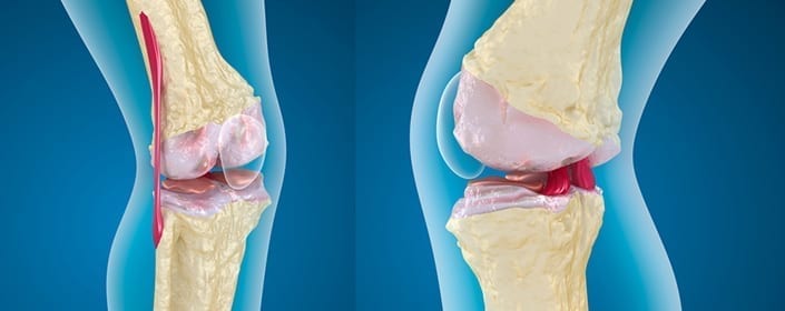
Given that many cartilage defects do not tend to repair themselves and can often lead to osteoarthritis, it is important to find effective ways to regenerate cartilage. There are a number of limitations to the current therapies, but this study demonstrates how our tools for cartilage repair are progressing and the promise of stem cells to help rebuild defective cartilage.
Learn more about using stem cells for orthopedics here.
Reference
Itokazu et al. (2016). Transplantation of scaffold-free cartilage-like cell-sheets made from human bone marrow mesenchymal stem cells for cartilage repair. Cartilage, 7(4), 361-372.
 St. Petersburg, Florida