by admin | Jun 12, 2017 | Studies
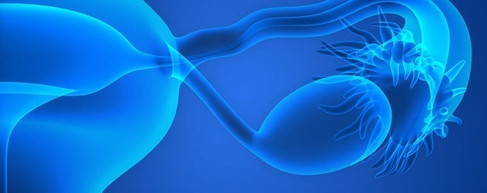
One of the several unfortunate side effects of chemotherapy, which is often used to combat cancer, is damage to the ovaries. Researchers have recently shown that the use of human umbilical cord mesenchymal stem cells provides one potentially promising way to treat the ovaries after the damage has occurred.
Depending on the age of the patient, the impact of chemotherapy-induced damage to the ovaries on quality of life can range from mild to devastating. For young patients who hope to have children, it is important to restore ovary function. Stem cells may provide a way to achieve this goal.
In the current study, researchers showed a number of positive effects of both injecting stem cells directing into the ovary, as well as injecting them less invasively, outside the body. The stem cell treatments were associated with recovery of the estrous cycle, and they led to a rise in sex hormone levels. In some cases, fertility was even restored and led to offspring that appeared to develop normally.
Though the restoration of ovary function occurred faster when the stem cells were injected into the ovaries, the long-term results of the two strategies were similar. These results suggest that a non-invasive form of stem cell therapy could be effective. However, when cells were injected directly into the ovary, they distributed within the ovary and uterus, whereas those injected from outside the body reached not only the ovary and uterus, but also the kidney, liver, and lungs. From this standpoint, direct injection of stem cells into the ovaries may be more desirable than injection from outside the body. However,
These data represent early evidence for the ability of stem cells to help reverse some of the damage that ovaries endure due to chemotherapy. However, the observation that ovary function can be improved as a result of stem cell injection provides an opportunity for further exploration into potential treatments for women whose ovaries have been damaged by chemotherapy.
Learn more about why umbilical cord stem cells are showing promise for stem cell-based therapies here.
Reference
Zhu et a l. (2015). Human umbilical cord mesenchymal stem cell transplantation restores damaged ovaries. Journal of Cellular & Molecular Medicine, 19(9), 2108-2117.
by admin | Jun 1, 2017 | Studies
Muscle atrophy refers to the wasting of muscles, which can occur due to lack of exercise or as a result of disease or even medication. Some forms of muscle atrophy can be treated with changes to the diet, exercise, or physical therapy, whereas others can be treated with clinical interventions like ultrasound therapy or surgery. However, researchers have now shown that stem cells may provide a way to prevent muscle atrophy in the first place.
While there may be visible signs of muscle atrophy, there are certain physiological events that allow doctors and scientists to determine with a greater degree of certainty whether muscle atrophy is occurring. For instance, when muscle atrophy occurs, certain proteins are expressed in lower amounts than they would normally be. The existence of these proteins gives researchers the opportunity to measure the levels of these proteins and thereby determine if and to what extent muscles are atrophying.
In the current study, researchers showed that in the presence of dexamethasone, a compound known to induce muscle atrophy, a mesenchymal stem cell-conditioned medium minimized the extent of atrophy. The researchers reached this conclusion because the expression of muscle atrophy-related protein was closer to normal when the mesenchymal stem cell medium was present than when it was not.
This preliminary evidence suggests that stem cells may be able to prevent muscle wasting and atrophy. Further studies that examine the impact of stem cells on muscle function will further our understanding of if and how stem cells will be able to be leveraged for the development of treatments or preventions for muscle atrophy.
Learn more about five benefits of stem cell therapy here.
Reference
Park et al. (2016). Umbilical cord mesenchymal stem cell-conditioned media prevent muscle atrophy by suppressing muscle atrophy-related proteins and ROS generation. In Vitro Cellular and Developmental Biology – Animal, 52(1), 68-76.
by admin | May 28, 2017 | Studies
Diabetic kidney disease often occurs in those with diabetes mellitus, even when they are undergoing normal diabetes treatment. Given the success that has been seen with a number of stem cell therapies and the lack of highly effective treatment options for diabetic kidney disease, much effort has been focused on determining if and how stem cells could be applied to this disease. In a recent review published in Current Diabetic Reports, researchers reviewed the progress that has occurred thus far in the work toward a potential stem cell therapy for diabetic kidney disease.
Diabetic kidney disease is complex because it involves problems both within the kidney, as well as more systemic issues that arise throughout the body. Because of its complexity, effective treatment is challenging. One way the disease has been treated is by inhibiting a hormone system, called the renin-angiotensin-aldosterone system, which regulates blood pressure in the arteries and the concentration of plasma sodium. When the system is too active, blood pressure increases, so inhibiting the system can help to lower that blood pressure. However, drugs for targeting other features of diabetic kidney disease have not yet proved to be significantly useful.
The type of stem cell that is particularly promising for use in diabetic kidney disease is the mesenchymal stem cell. Mesenchymal stem cells are easy to access and inherently possess some of the characteristics that would prove useful in treating kidney disease, including working to reduce inflammation protect cells. Preclinical studies have shown promising results in using mesenchymal stem cells to slow the progression of diabetic kidney disease. Clinical trials are currently underway to help determine if these cells can indeed help those with diabetic kidney disease and to additionally help to develop specific protocol for applying stem cell treatments in this disease.
Meanwhile, basic science research continues to be undertaken to help elucidate the mechanisms by which stem cells may impart benefits on those with diabetic kidney disease. Understanding the mechanism of action could help inform the development of specific therapies and efficiently end routes of investigation that are unlikely to be fruitful.
Studies have shown that stem cells can be useful for treating kidney disease. Read about it here.
Reference
Griffin, TP et al (2016). The promise of mesenchymal stem cell therapy for diabetic kidney disease. Current Diabetic Reports, 16(5), 42.
by admin | May 5, 2017 | Studies
Patients with multiple sclerosis suffer from an impairment in the function of specific cells of their immune system, known as T regulatory cells. The cause for the disease is not clear, and though treatments do exist, they tend to be expensive and to also carry the risk for toxic effects. To overcome the limitations of current treatment options, researchers have begun to explore the use of stem cells in the development of new treatments.
In a study recently published in the journal Oncotarget, researchers described the preliminary results of a study aimed at identifying the feasibility of using stem cells to improve the functioning of T regulatory cells in those with multiple sclerosis. Umbilical cord-derived mesenchymal stem cells were the chosen cell type for the experiment because this specific type of stem cell has been shown to affect the functioning of immune cells.
The researchers confirmed the idea that T regulatory cells are severely impaired in multiple sclerosis and were able to show that umbilical cord-derived mesenchymal stem cells could recover the functioning of the T regulatory cells of multiple sclerosis patients. Not only were there more living, active T regulatory cells in conditions that included the stem cells versus those without stem cells, but these cells also demonstrated the normal types of activities that T regulatory cells contribute to the immune system.
Previous research has established the potential for targeting T regulatory cells in the treatment of multiple sclerosis, but these studies have been conducted primarily in animal models of the disease. These newer results are the first to demonstrate the impact of umbilical cord-derived mesenchymal stem cells on immune cells of patients with multiple sclerosis.
Read more about how adult stem cell therapy can assist in the reversal of challenging symptoms and damage associated with MS here.
Reference
Yang, H. et al. (2016). Umbilical cord-derived mesenchymal stem cells reversed the suppressive deficiency of T regulatory cells from peripheral blood of patients with multiple sclerosis in co-culture – a preliminary study. Oncotarget, 7: 72537-72545.
by admin | Apr 20, 2017 | Studies
Given the promise they have shown for treating neurodegenerative disease and different types of damage to the brain, it is likely that stem cells have the potential to combat hearing loss. Researchers have recently shown how combining neural stem cells with a specific compound, called morin hydrate, can protect against hearing loss.
Unlike cells of many other organs, hair cells in the inner ear, referred to as cochlear hair cells, are not able self-renew once they are damaged. As a result, damage to these cells of the inner ear increase vulnerability to hearing loss. Replacing these cells with stem cells, has therefore been thought to be a good way to try to reduce this vulnerability.
A major challenge in using stem cells to combat disease or disorders such as hearing loss is finding the best way to transplant the cells so that they survive, differentiate, and function. In this study, published last month in the Journal of Cellular and Molecular Medicine, scientists set out to determine if combining neural stem cells with morin hydrate may make those stem cells more viable and increase their ability to help with hearing loss. Morin hydrate was chosen because it has been associated with a number of beneficial effects, including the suppression of inflammation and cancer activity.
The results of their study showed that morin hydrate was able to improve neural stem cell survival and proliferation and that the compound enabled the types of connections between cells that are necessary for proper neuronal functioning and the protection against hearing loss. In addition to the positive impact of morin hydrate on the stem cells themselves, the compound was also shown to prevent hearing loss that resulted from gentamicin, a specific type of antibiotic.
These initial results demonstrate that the inclusion of morin hydrate in stem cell therapy has promise. Future research will help determine how the combination of morin hydrate and stem cells could be used to address hearing loss and potentially other disorders.
Learn how stem cells can help reduce noise-induced hearing loss here.
Reference
He, Q., Jia, Z., Zhang, Y., & Ren, X. (2017). Morin hydrate promotes inner ear neural stem cell survival and differentiation and protects cochlea against neuronal hearing loss. Journal of Cellular & Molecular Medicine, 21(3), 600-608.
by admin | Apr 14, 2017 | Studies
When cochlear cells within the ear are damaged from exposure to high levels of noise, long-term, permanent hearing loss can occur. Because this hearing loss is associated with damaged cells, researchers have reasoned that replacing those cells with stem cells may provide a means for reversing noise-induced hearing loss. A recent study, published in the journal Neurobiological Disorders has shown that the transplant of epithelial stem cells can in fact help with this type of hearing loss.
The stem cells that were used in the study were isolated from the tongue and were shown to have the ability to survive and proliferate outside the body. Once transplanted, they were also shown to survive and to integrate themselves appropriately.
The auditory brainstem response (ABR) threshold test was used before and after transplantation to determine whether the stem cells actually impacted hearing loss level. The ABR test is a neurological test that assesses whether the brainstem responds to auditory clicking sounds. The test was developed in 1971 and is now the most widely used test for evaluating responses to auditory stimuli. The test reveals the threshold at which noise can produce a response, with lower thresholds indicating better auditory functioning.
Compared to before the transplantation, tests performed 4 weeks after transplantation showed that the stem cell transplants were associated with lower ABR thresholds. Thus, not only did the stem cells survive, proliferate, and integrate normally within the ear, but they were also associated with improved auditory abilities.
These results indicate that stem cells are a promising candidate for reversing long-term hearing loss that is caused by noise-induced damage to cells of the inner ear. Further research will help to clarify the best ways these cells may be used to reverse hearing loss and to what extent their application can benefit those who have suffered noise-induced hearing loss. It is also possible that the relevant research will help reveal ways that stem cells can be used to help those who suffer from other types of hearing loss as well.
Read more about how stem cells treated with morin hydrate can protect against hearing loss here.
Reference
Sullivan, J.M., Cohen, M.A., Pandit, S.R., Sahota, R.S., Borecki, A.A., & Oleskevich, S. (2011). Effect of epithelial stem cell transplantation on noise-induced hearing loss in adult mice. Neurobiological Disorders. 41(2), 552-559.


 St. Petersburg, Florida